HIV: neurocomplications
Focal neurological lesions
Toxoplasmosis
- accounts for around 50% of cerebral lesions in patients with HIV
- constitutional symptoms, headache, confusion, drowsiness
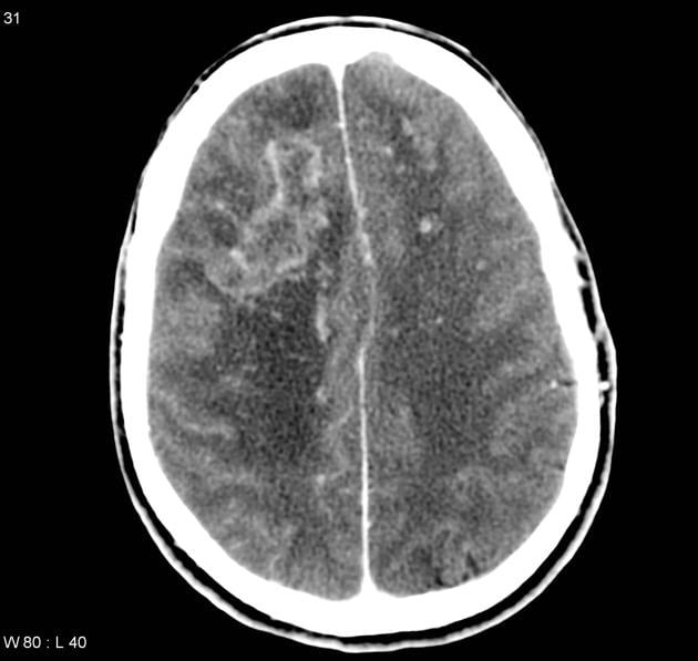
- CT: usually single or multiple ring enhancing lesions, mass effect may be seen
- management: sulfadiazine and pyrimethamine
 | |
| © Image used on license from Radiopaedia | |
Cerebral toxoplasmosis: CT scan with contrast showing multiple ring enhancing lesions
 | |
| © Image used on license from Radiopaedia | |
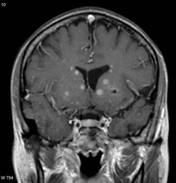
Cerebral toxoplasmosis: MRI (T1 C+) demonstrates multiple small peripherally enhancing nodules located predominantly in the basal ganglia as well as the central portions of the cerebellar hemispheres. Only a small amount of surrounding oedema is present.
Primary CNS lymphoma
- accounts for around 30% of cerebral lesions
- associated with the Epstein-Barr virus
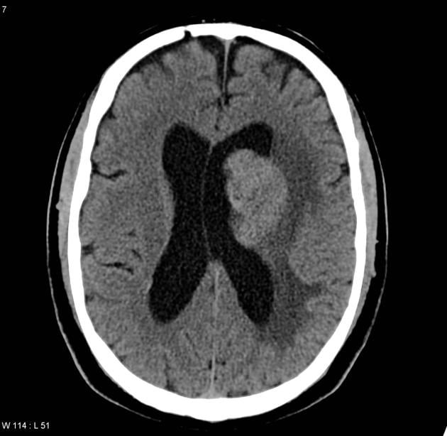
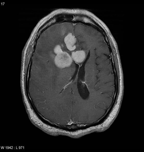
- CT: single or multiple homogenous enhancing lesions
- treatment generally involves steroids (may significantly reduce tumour size), chemotherapy (e.g. methotrexate) + with or without whole brain irradiation. Surgical may be considered for lower grade tumours
 | |
| © Image used on license from Radiopaedia | |
Primary CNS lymphoma: Non-contrast CT demonstrates a hyper-attenuating mass adjacent to the left lateral ventricle, with no calcification or haemorrhage.
 | |
| © Image used on license from Radiopaedia | |
Primary CNS lymphoma: MRI (T1 C+) demonstrates a large multilobulated mass in the right frontal lobe. It homogeneously enhances and extends to involve the caudate and the periventricular area. There is significant mass effect.
Differentiating between toxoplasmosis and lymphoma is a common clinical scenario in HIV patients. It is clearly important given the vastly different treatment strategies. The table below gives some general differences. Please see the Radiopaedia link for more details.
| Toxoplasmosis | Lymphoma |
|---|---|
| Multiple lesions Ring or nodular enhancement Thallium SPECT negative | Single lesion Solid (homogenous) enhancement Thallium SPECT positive |
Tuberculosis
- much less common than toxoplasmosis or primary CNS lymphoma
- CT: single enhancing lesion
Generalised neurological disease
Encephalitis
- may be due to CMV or HIV itself
- HSV encephalitis but is relatively rare in the context of HIV
- CT: oedematous brain
Cryptococcus
- most common fungal infection of CNS
- headache, fever, malaise, nausea/vomiting, seizures, focal neurological deficit
- CSF: high opening pressure, India ink test positive
- CT: meningeal enhancement, cerebral oedema
- meningitis is typical presentation but may occasionally cause a space occupying lesion
Progressive multifocal leukoencephalopathy (PML)
- widespread demyelination
- due to infection of oligodendrocytes by JC virus (a polyoma DNA virus)
- symptoms, subacute onset : behavioural changes, speech, motor, visual impairment
- CT: single or multiple lesions, no mass effect, don't usually enhance. MRI is better - high-signal demyelinating white matter lesions are seen
AIDS dementia complex
- caused by HIV virus itself
- symptoms: behavioural changes, motor impairment
- CT: cortical and subcortical atrophy