Acute kidney injury: a very basic introduction
On this page
Acute kidney injury (AKI), previously termed acute renal failure, describes a reduction in renal function following an insult to the kidneys. In years gone by the kidneys were very much a neglected organ in acute medicine - the recognition of decreasing renal function was often slow and action limited. Around 15% of patients admitted to hospital develop AKI.
Whilst most patients with AKI recover their renal function there are many patients who will have long term impaired kidney function due to AKI. As well as long-term morbidity, AKI may also result in acute complications including death. Whilst exact figures are difficult to calculate NICE estimate that inpatient mortality of AKI in the UK might typically be 25-30% or more.
Causes of AKI are traditionally divided into prerenal, intrinsic and postrenal causes
Think of what causes big problems in other major organs. In the heart a lack of blood (ischaemia) to the myocardium causes a myocardial infarction. In a similar fashion 85% of strokes are causes be ischaemia to the brain. The same goes for the kidneys. One of the major causes of AKI is ischaemia, or lack of blood flowing to the kidneys.
Examples
Intrinsic
The second group of causes relate to intrinsic damage to the glomeruli, renal tubules or interstitium of the kidneys themselves. This may be due to toxins (drugs, contrast etc) or immune-mediated glomuleronephritis.
Examples
Postrenal
The third group relates to problems after the kidneys. This is where there is an obstruction to the urine coming from the kidneys resulting in things 'backing-up' and affecting the normal renal function. An example could be a unilateral ureteric stone or bilateral hydroneprosis secondary to acute urinary retention caused by benign prostatic hyperplasia.
Examples
One of the keys to reducing the incidence of AKI is identifying patient who are at increased risk. NICE support this approach and have published guidelines suggesting which patients are at greater risk.
Risk factors for AKI include:
Preventing AKI
By identifying patients at increased risk of AKI (see above) it may be possible to take steps to reduce the risk. For example, patients who are at risk of AKI and who are undergoing an investigation requiring contrast are usually given IV fluids to reduce the risk. Certain drugs such as ACE inhibitors and ARBs may also be temporarily stopped.
It's best to work backwards and think about what kidneys actually do. The kidneys are primarily responsible for fluid balance and maintaining homeostasis. Therefore two of the key ways AKI may be detected are:
Symptoms and signs
Many patients with early AKI may experience no symptoms. However, as renal failure progresses the following may be seen:
Detection
One of the most common blood tests performed on the wards is 'urea and electrolytes' or 'U&Es'. This returns a number of markers, including
NICE recommend that we can use a variety of different criteria to make an official diagnosis of AKI. They state:
Detect acute kidney injury, in line with the (p)RIFLE, AKIN or KDIGO definitions, by using any of the following criteria:
Urinanalysis
Imaging
The management of AKI is largely supportive. This means patients require careful fluid balance to ensure that the kidneys are properly perfused but not excessively to avoid fluid overload. It is also important to review a patient's medication list to see what treatments may either be exacerbating their renal dysfunction or may be dangerous as a consequence of renal dysfunction. The table below gives some examples of common drugs:
Treatments which are not recommend include the routine use of loop diuretics (to artificially boost urine output) and low-dose dopamine (in an attempt to increase renal perfusion). There is however a role for loop diuretics in patients who experience significant fluid overload.
Hyperkalaemia also needs prompt treatment to avoid arrhythmias which may potentially be life-threatening. The table below categorises the different treatments for hyperkalaemia:
Specialist input from a nephrologist is required for cases where the cause is not known or where the AKI is severe.
All patients with suspected AKI secondary to urinary obstruction require prompt review by a urologist.
Renal replacement therapy (e.g. haemodialysis) is used when a patient is not responding to medical treatment of complications, for example hyperkalaemia, acidosis or uraemia.
Whilst most patients with AKI recover their renal function there are many patients who will have long term impaired kidney function due to AKI. As well as long-term morbidity, AKI may also result in acute complications including death. Whilst exact figures are difficult to calculate NICE estimate that inpatient mortality of AKI in the UK might typically be 25-30% or more.
 | |
| © Image used on license from PathoPic | |
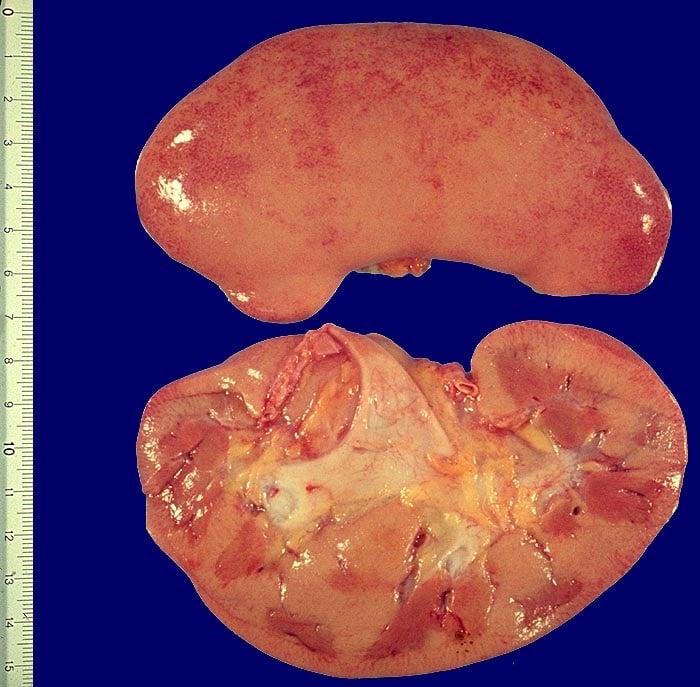
Specimen from a patient who had an acute kidney injury. Note the marked pallor of the cortex in certain areas, contrasting to the darker areas of surviving medullary tissue.
What causes AKI?
Causes of AKI are traditionally divided into prerenal, intrinsic and postrenal causes
Prerenal
Think of what causes big problems in other major organs. In the heart a lack of blood (ischaemia) to the myocardium causes a myocardial infarction. In a similar fashion 85% of strokes are causes be ischaemia to the brain. The same goes for the kidneys. One of the major causes of AKI is ischaemia, or lack of blood flowing to the kidneys.
Examples
- hypovolaemia secondary to diarrhoea/vomiting
- renal artery stenosis
Intrinsic
The second group of causes relate to intrinsic damage to the glomeruli, renal tubules or interstitium of the kidneys themselves. This may be due to toxins (drugs, contrast etc) or immune-mediated glomuleronephritis.
Examples
- glomerulonephritis
- acute tubular necrosis (ATN)
- acute interstitial nephritis (AIN), respectively
- rhabdomyolysis
- tumour lysis syndrome
Postrenal
The third group relates to problems after the kidneys. This is where there is an obstruction to the urine coming from the kidneys resulting in things 'backing-up' and affecting the normal renal function. An example could be a unilateral ureteric stone or bilateral hydroneprosis secondary to acute urinary retention caused by benign prostatic hyperplasia.
Examples
- kidney stone in ureter or bladder
- benign prostatic hyperplasia
- external compression of the ureter
 | |
| © Image used on license from PathoPic | |
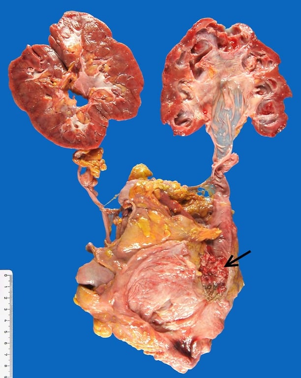
This patient had an invasive papillary tumour of the distal left ureter, indicated by the arrow. This resulted in the ureter becoming blocked, resulting in a left hydroureter and hydronephrosis. Note the thinning of the renal cortex of the left kidney compared to the right.
Who is at an increased risk of AKI?
One of the keys to reducing the incidence of AKI is identifying patient who are at increased risk. NICE support this approach and have published guidelines suggesting which patients are at greater risk.
Risk factors for AKI include:
- chronic kidney disease
- other organ failure/chronic disease e.g. heart failure, liver disease, diabetes mellitus
- history of acute kidney injury
- use of drugs with nephrotoxic potential (e.g. NSAIDs, aminoglycosides, ACE inhibitors, angiotensin II receptor antagonists [ARBs] and diuretics) within the past week
- use of iodinated contrast agents within the past week
- age 65 years or over
- neurological or cognitive impairment or disability, which may mean limited access to fluids because of reliance on a carer
Preventing AKI
By identifying patients at increased risk of AKI (see above) it may be possible to take steps to reduce the risk. For example, patients who are at risk of AKI and who are undergoing an investigation requiring contrast are usually given IV fluids to reduce the risk. Certain drugs such as ACE inhibitors and ARBs may also be temporarily stopped.
What happens when kidneys stop working?
It's best to work backwards and think about what kidneys actually do. The kidneys are primarily responsible for fluid balance and maintaining homeostasis. Therefore two of the key ways AKI may be detected are:
- a reduced urine output. This is termed oliguria and is defined as a urine output of less than 0.5 ml/kg/hour
- fluid overload
- a rise in molecules that the kidney normal excretes/maintains a careful balance of. Examples include potassium, urea and creatinine
Symptoms and signs
Many patients with early AKI may experience no symptoms. However, as renal failure progresses the following may be seen:
- reduced urine output
- pulmonary and peripheral oedema
- arrhythmias (secondary to changes in potassium and acid-base balance)
- features of uraemia (for example, pericarditis or encephalopathy)
Detection
One of the most common blood tests performed on the wards is 'urea and electrolytes' or 'U&Es'. This returns a number of markers, including
- sodium
- potassium
- urea
- creatinine
NICE recommend that we can use a variety of different criteria to make an official diagnosis of AKI. They state:
Detect acute kidney injury, in line with the (p)RIFLE, AKIN or KDIGO definitions, by using any of the following criteria:
- a rise in serum creatinine of 26 micromol/litre or greater within 48 hours
- a 50% or greater rise in serum creatinine known or presumed to have occurred within the past 7 days
- a fall in urine output to less than 0.5 ml/kg/hour for more than 6 hours in adults and more than
Urinanalysis
- all patients with suspected AKI should have urinanalysis
Imaging
- if patients have no identifiable cause for the deterioration or are at risk of urinary tract obstruction they should have a renal ultrasound within 24 hours of assessment.
Management
The management of AKI is largely supportive. This means patients require careful fluid balance to ensure that the kidneys are properly perfused but not excessively to avoid fluid overload. It is also important to review a patient's medication list to see what treatments may either be exacerbating their renal dysfunction or may be dangerous as a consequence of renal dysfunction. The table below gives some examples of common drugs:
| Usually safe to continue in AKI | Should be stopped in AKI as may worsen renal function | May have to be stopped in AKI as increased risk of toxicity (but doesn't usually worsen AKI itself) |
|---|---|---|
| • Paracetamol • Warfarin • Statins • Aspirin (at a cardioprotective dose of 75mg od) • Clopidogrel • Beta-blockers | • NSAIDs • Aminoglycosides • ACE inhibitors • Angiotensin II receptor antagonists • Diuretics | • Metformin • Lithium • Digoxin |
Treatments which are not recommend include the routine use of loop diuretics (to artificially boost urine output) and low-dose dopamine (in an attempt to increase renal perfusion). There is however a role for loop diuretics in patients who experience significant fluid overload.
Hyperkalaemia also needs prompt treatment to avoid arrhythmias which may potentially be life-threatening. The table below categorises the different treatments for hyperkalaemia:
| Stabilisation of the cardiac membrane | • Short-term shift in potassium from extracellular to intracellular fluid compartment | • Removal of potassium from the body |
|---|---|---|
| • Intravenous calcium gluconate | • Combined insulin/dextrose infusion • Nebulised salbutamol | • Calcium resonium (orally or enema) • Loop diuretics • Dialysis |
Specialist input from a nephrologist is required for cases where the cause is not known or where the AKI is severe.
All patients with suspected AKI secondary to urinary obstruction require prompt review by a urologist.
Renal replacement therapy (e.g. haemodialysis) is used when a patient is not responding to medical treatment of complications, for example hyperkalaemia, acidosis or uraemia.