Action Potential
On this page
An action potential (or nerve impulse) occurs when the membrane potential within a cell rapidly rises and falls. This is termed depolarisation. The depolarisation results in adjacent cells depolarising. Action potentials only occur in excitable cells which include neurones, myocytes and endocrine cells.
Cells contain negatively charged proteins that are unable to cross the cell membrane. These negatively charged molecules attract positively charged ions.
This forces K+ ions into the cell. The subsequent K+ concentration gradient drives K+ out of the cell i.e there is an electrical gradient and a concentration gradient that drive K+ in opposing directions. These two forces reach an equilibrium where there is a relative negative charge within the cell compared to the outside i.e. a potential difference exists.
It would be expected that other positively charged ions such as Na+ would act in the same way as K+ but they do not due to differences in permeability whilst a cell is at rest (no action potential being generated or transmitted).
Potassium permeability (Pk) is much higher in cell membranes than other ions e.g. potassium permeability is 25-100 times higher than sodium permeability (PNa) in excitable tissues (nerve and muscle cells) at rest. The result is that resting membrane potential (Em) is very close to K+ equilibrium potential (Ek). Some Na+ does leak into the cell along its electrochemical gradient but this is then actively pumped across the cell membrane to maintain a large Na+ concentration gradient.
As mentioned above sodium permeability (PNa) is low at rest but if PNa suddenly increases and becomes greater than potassium permeability (Pk) the membrane potential shifts form being close to K+ equilibrium potential (Ek) to being close to Na+ equilibrium potential (ENa). This is what happens during an action potential when Na+ channels are opened causing a sudden rise in sodium permeability where PNa becomes 10 times that of Pk
In nerve and skeletal muscle action potentials are initiated via the opening of ligand gated Na+ channels by neurotransmitters. The increased sodium permeability allows sodium ions to flow into the cell and causes the membrane potential to become more positive. If the stimulus is strong enough and causes the membrane potential to meet the threshold potential voltage gated Na+ channels then open. The opening of voltage gated Na+ channels promotes further voltage gated Na+ channels to open and rapid depolarisation to occur. The depolarisation ends when the positive voltage inactivates the voltage gated Na+ channels.
As the voltage gated Na+ channels close, voltage gated K+ channels open and potassium flows out of the cell. Delayed closure of these potassium channels results in a transient hyperpolarisation. Following depolarisation the sodium channels remain inactive for around 1ms and for a period cannot depolarise regardless of the level of stimulus, this is known as the absolute refractory period. For a further 2-3ms whilst the cells are hyperpolarised they may be depolarised but require a larger stimulus, this is known as the relative refractory period.
At the end of repolarization the cell has essentially swapped intracellular K+ for Na+ as it has had a big influx of sodium followed by an efflux of potassium. The ionic concentrations are restored via the sodium pump (AKA the Na+-K+-ATPase).
Once triggered action potentials will travel throughout the excitable cell i.e. it is propagated. Action potentials are always the same amplitude and therefore said to be
These differences include:
Membrane potential
A potential difference exists across the membranes of all cells. Excitable tissues can generate action potentials which are transient depolarisations of a cell as a result of ion movement. The inside of a cell membrane is negatively charged compared to the outside of the cell membrane. In excitable tissues the resting membrane potential is -60 to -90mV.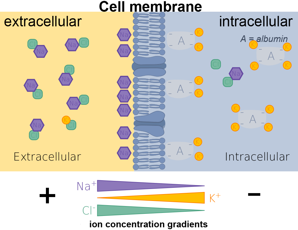 | |
click on image to enlarge
Cells contain negatively charged proteins that are unable to cross the cell membrane. These negatively charged molecules attract positively charged ions.
This forces K+ ions into the cell. The subsequent K+ concentration gradient drives K+ out of the cell i.e there is an electrical gradient and a concentration gradient that drive K+ in opposing directions. These two forces reach an equilibrium where there is a relative negative charge within the cell compared to the outside i.e. a potential difference exists.
The potential difference across the cell membrane is the result of an equilibrium between electrical gradient and concentration gradient.
It would be expected that other positively charged ions such as Na+ would act in the same way as K+ but they do not due to differences in permeability whilst a cell is at rest (no action potential being generated or transmitted).
Potassium permeability (Pk) is much higher in cell membranes than other ions e.g. potassium permeability is 25-100 times higher than sodium permeability (PNa) in excitable tissues (nerve and muscle cells) at rest. The result is that resting membrane potential (Em) is very close to K+ equilibrium potential (Ek). Some Na+ does leak into the cell along its electrochemical gradient but this is then actively pumped across the cell membrane to maintain a large Na+ concentration gradient.
Resting membrane potentials are the result of the asymmetric distributions of Na+ and K+.
As mentioned above sodium permeability (PNa) is low at rest but if PNa suddenly increases and becomes greater than potassium permeability (Pk) the membrane potential shifts form being close to K+ equilibrium potential (Ek) to being close to Na+ equilibrium potential (ENa). This is what happens during an action potential when Na+ channels are opened causing a sudden rise in sodium permeability where PNa becomes 10 times that of Pk
The action potential
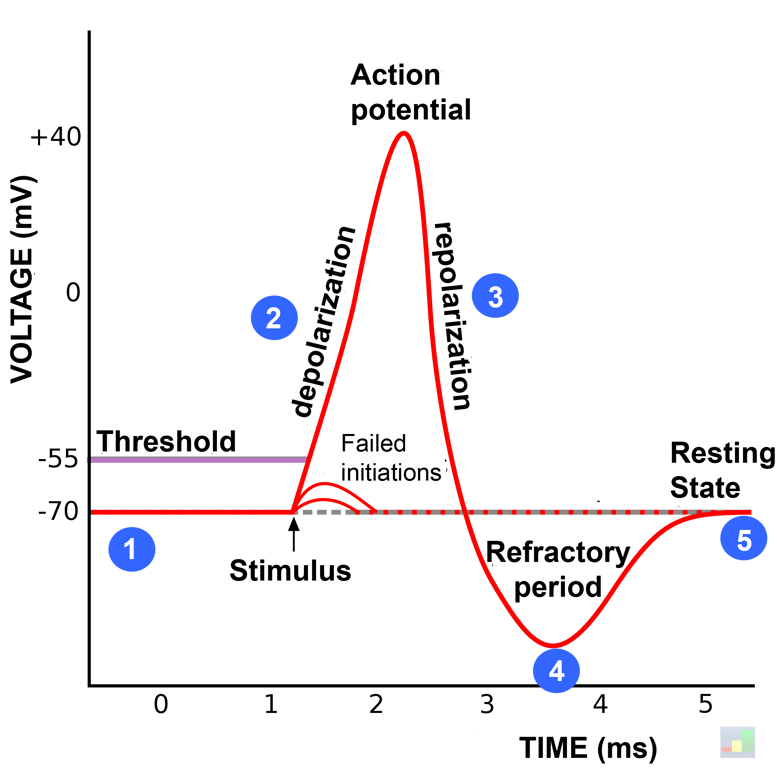 | |
Action potential for a typical neurone. At the resting state (1) the neurone has a net negative charge inside. Once the action potential is triggered, a stimulus triggers depolarisation (2) where sodium channels, open allowing sodium ions to pass rapidly through the cell membrane along their concentration gradient into the cell, resulting in a net positive charge inside the neurone relative to the extracellular fluid. After the action potential peak is reached, the neurone begins repolarisation (3), where sodium channels close and potassium channels open, allowing potassium ions to cross the membrane into the extracellular fluid, returning the membrane potential to a negative value. Finally, there is a refractory period (4), during which the voltage-dependent ion channels are inactivated and Na+ and K+ ions are exchanged to return them to their resting state ion distributions across the membrane (5), and the neurone is ready to repeat the process for the next action potential. Click on image to enlarge
In nerve and skeletal muscle action potentials are initiated via the opening of ligand gated Na+ channels by neurotransmitters. The increased sodium permeability allows sodium ions to flow into the cell and causes the membrane potential to become more positive. If the stimulus is strong enough and causes the membrane potential to meet the threshold potential voltage gated Na+ channels then open. The opening of voltage gated Na+ channels promotes further voltage gated Na+ channels to open and rapid depolarisation to occur. The depolarisation ends when the positive voltage inactivates the voltage gated Na+ channels.
As the voltage gated Na+ channels close, voltage gated K+ channels open and potassium flows out of the cell. Delayed closure of these potassium channels results in a transient hyperpolarisation. Following depolarisation the sodium channels remain inactive for around 1ms and for a period cannot depolarise regardless of the level of stimulus, this is known as the absolute refractory period. For a further 2-3ms whilst the cells are hyperpolarised they may be depolarised but require a larger stimulus, this is known as the relative refractory period.
At the end of repolarization the cell has essentially swapped intracellular K+ for Na+ as it has had a big influx of sodium followed by an efflux of potassium. The ionic concentrations are restored via the sodium pump (AKA the Na+-K+-ATPase).
Once triggered action potentials will travel throughout the excitable cell i.e. it is propagated. Action potentials are always the same amplitude and therefore said to be
all or nothing
Cardiac myocytes & pacemaker cells
Cardiac cells have some unique differences to other excitable cells such as skeletal muscle and neurones. There are also difference between cardiac pacemaker cells and cardiac myocytes.These differences include:
- Resting potential value
- Threshold potential value
- Action potential profile variations due to different ion channels utilised for depolarisation and repolarization as shown by the images below:
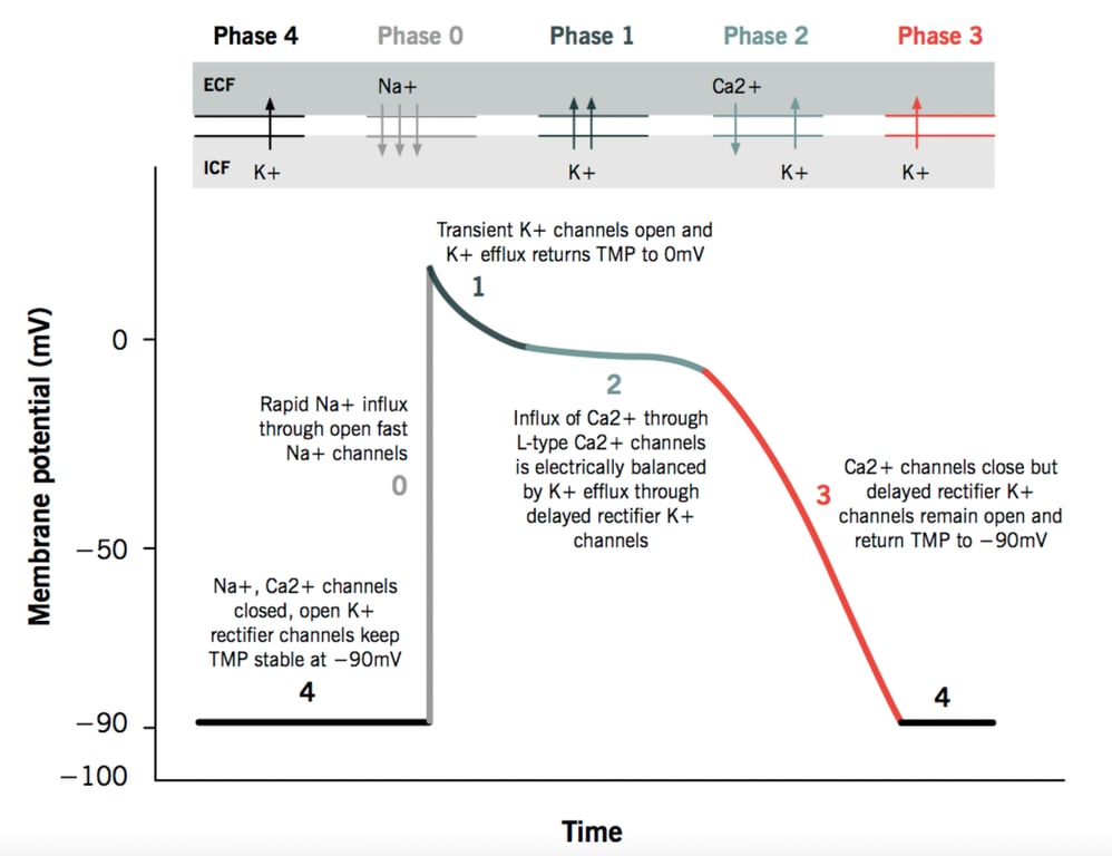 | |
Cardiac myocyte action potential. Click on image to enlarge
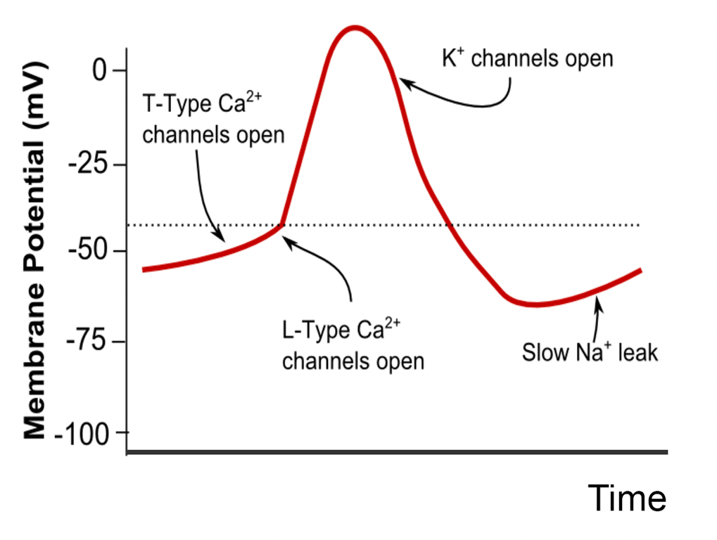 | |
Pacemaker action potential. Click on image to enlarge