Adrenal function
On this page
The adrenal glands are paired endocrine organs located on the superior pole of each kidney.
The adrenals are retroperitoneal, with parietal peritoneum covering their anterior surface only. Perinephric fascia encloses the adrenal glands (and the kidneys) and attaches them to the crura of the diaphragm. A layer of perirenal fat separates the kidneys from the adrenals.
The adrenal gland is divided into two functionally active and distinct regions:
Adrenal Cortex
The adrenal cortex is the outermost functional layer and is divided into three regions (from superficial to deep):
Adrenaline accounts for around 80% of catecholamine secretion and noradrenaline 20%. Catecholamines play important roles in normal physiological functioning such as maintenance of blood pressure but their role is enhanced by stress.
The adrenal medulla is functionally part of the sympathetic nervous system. Catecholamine release is stimulated by sympathetic preganglionic neurones located in the spinal cord. Catecholamines act on guanosine triphosphate binding protein (G-protein) coupled adrenoreceptors. The action depends on the class of adrenoreceptor (α1, α2, β1, β2 or β3) but the response to catecholamine stimulation is the same as sympathetic nerve stimulation.
In stress responses the above catecholamine mediated actions support increased physical activity. At normal plasma concentrations adrenaline only stimulates β adrenoreceptors but at elevated levels α adrenreceptors are stimulated.
*At low plasma levels, adrenaline preferentially binds to high affinity β2 adrenoceptors and causes vasodilation which results in a drop in systemic vascular resistance (SVR). At increased adrenaline levels, lower affinity α-adrenoceptors begin binding adrenaline and the balance of vasodilatory and vasoconstrictor actions of epinephrine shifts to net vasoconstriction (increased systemic vascular resistance).
The breakdown may then proceed by a variety of pathways producing the following metabolites that are secreted in the urine.
The primary function of the adrenal glands is to secrete steroid and catecholamine hormones into the blood
The adrenals are retroperitoneal, with parietal peritoneum covering their anterior surface only. Perinephric fascia encloses the adrenal glands (and the kidneys) and attaches them to the crura of the diaphragm. A layer of perirenal fat separates the kidneys from the adrenals.
The adrenal gland is divided into two functionally active and distinct regions:
- Adrenal cortex
- Adrenal medulla
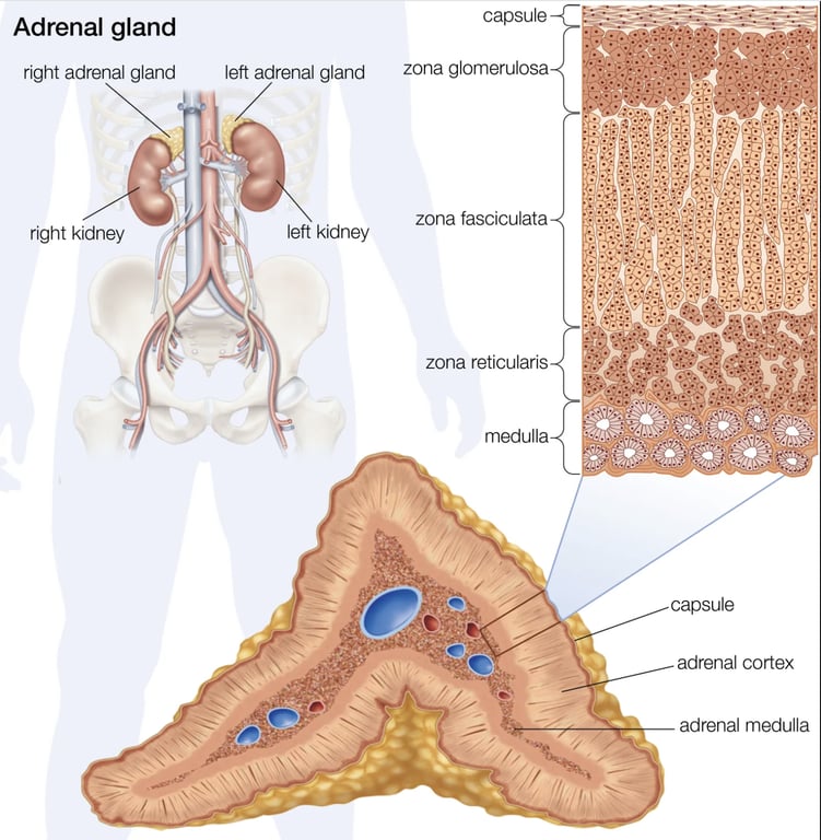 | |
Anatomy and histology of the adrenal glands. Click on image to enlarge
Adrenal Cortex
The adrenal cortex is the outermost functional layer and is divided into three regions (from superficial to deep):
- Zona glomerulosa: produces and secretes mineralocorticoids, aldosterone being the most important.
- Zona fasciculata: produces and secretes glucocorticoids, cortisol being the most important. Small amounts of androgens are also secreted by the zona fasciculata.
- Zona reticularis: produces and secretes androgens such as dehydroepiandrosterone (DHEA). Small amounts of corticosteroids are also secreted by the zona reticularis.
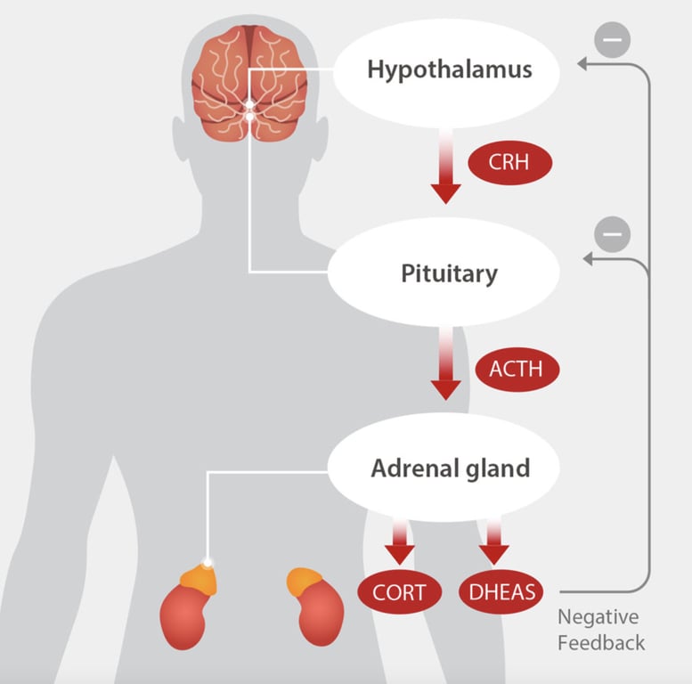 | |
Cortisol and DHEA secretion are stimulated by adrenocorticotropic (ACTH) hormone which in turn is released in response to corticotropin-releasing hormone (CRH) release from the hypothalamus. Click on image to enlarge
Metabolic and pharmacological effects of glucocorticoids (cortisol)
At high levels cortisol can mimic the action of aldosterone on the kidney resulting in Na+ ion retention and K+ ion loss.
Pharmacological effects of cortisol:
- Anti-inflammatory: cortisol upregulates the expression of anti-inflammatory proteins whilst downregulating the expression of pro-inflammatory proteins. Specifically cortisol inhibits production of interleukin 12 (IL-12), interferon gamma (IFN-gamma), IFN-alpha, and tumor necrosis factor alpha (TNF-alpha) by antigen-presenting cells (APCs) and T helper cells (Th1 cells). Cortisol upregulates the anti-inflammatory cytokines IL-4, IL-10, and IL-13 by Th2 cells.
- Weakened immune response: Cortisol inhibits proliferation of T-cells. Cortisol makes T-cells unresponsive to IL-1 which renders the T-cell unable to produce IL-2. Cortisol also downregulates the expression of IL-2 receptors on helper T-cells which are required to induce a Th1 'cellular' immune response.
Metabolic effects of cortisol:
- Increases glycolysis and gluconeogenesis in the liver (raising blood sugar)
- Increases insulin resistance
- Increase lipolysis
- Increases proteolysis (can lead to muscle wasting long term)
- Increases blood pressure via up-regulating alpha-1 receptors on arterioles
- Inhibits bone formation: decreases osteoblast activity, decreases type 1 collagen synthesis, decreases absorption of calcium from the gut & increases osteoclastic activity.
At high levels cortisol can mimic the action of aldosterone on the kidney resulting in Na+ ion retention and K+ ion loss.
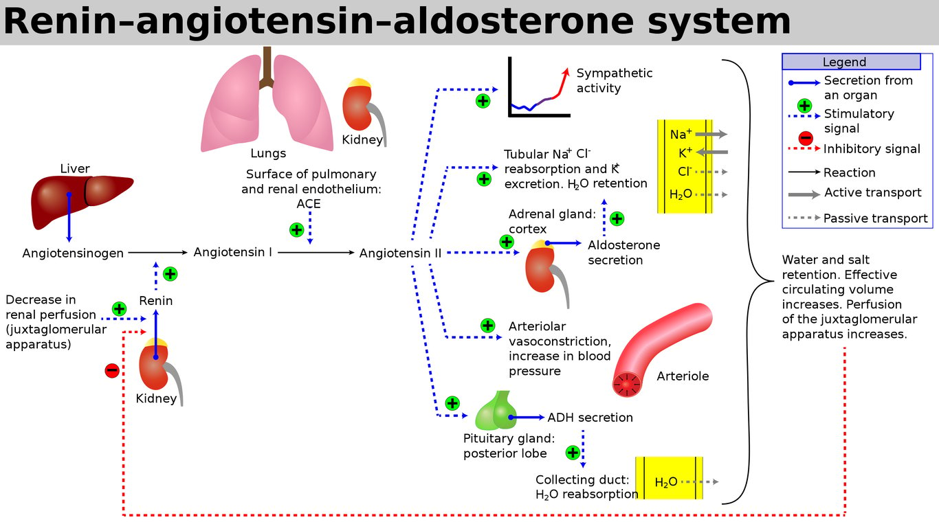 | |
Aldosterone secretion is regulated as per the renin-angiotensin system. Stimuli for aldosterone secretion are hypovolaemia, hyponatraemia or hyperkalaemia. Click on image to enlarge
Adrenal Medulla
The adrenal medulla contains chromaffin cells. Chromaffin cells synthesize and secrete the catecholamines adrenaline (epinephrine) and noradrenaline (norepinephrine) into the bloodstream. These catecholamines are synthesized from the amino acid tyrosine by several enzymatic steps that produce a series of metabolic intermediates such as dopamine.Adrenaline accounts for around 80% of catecholamine secretion and noradrenaline 20%. Catecholamines play important roles in normal physiological functioning such as maintenance of blood pressure but their role is enhanced by stress.
Stress stimulates catecholamine release.
The adrenal medulla is functionally part of the sympathetic nervous system. Catecholamine release is stimulated by sympathetic preganglionic neurones located in the spinal cord. Catecholamines act on guanosine triphosphate binding protein (G-protein) coupled adrenoreceptors. The action depends on the class of adrenoreceptor (α1, α2, β1, β2 or β3) but the response to catecholamine stimulation is the same as sympathetic nerve stimulation.
| Receptor | Response to stimulation |
|---|---|
| α1 | vasoconstriction |
| α2 | selective vasoconstriction, negative feedback inhibiting catecholamines |
| β1 | increased cardiac output |
| β2 or β3 | Increased glycolysis & lipolysis |
In stress responses the above catecholamine mediated actions support increased physical activity. At normal plasma concentrations adrenaline only stimulates β adrenoreceptors but at elevated levels α adrenreceptors are stimulated.
Adrenaline important effects summary
Cardiovascular effects
- Increased cardiac output
- Increased blood pressure
- Increased cardiac contractility
- Selective peripheral vasoconstriction of systemic arteries and veins
- Vasodilation in muscle and liver vasculatures
- Increased systemic vascular resistance at high doses*
- Bronchodilation
- Increased glycolysis
- Increased glycogenolysis
- Increased gluconeogenesis
- Increased lipolysis
*At low plasma levels, adrenaline preferentially binds to high affinity β2 adrenoceptors and causes vasodilation which results in a drop in systemic vascular resistance (SVR). At increased adrenaline levels, lower affinity α-adrenoceptors begin binding adrenaline and the balance of vasodilatory and vasoconstrictor actions of epinephrine shifts to net vasoconstriction (increased systemic vascular resistance).
Degradation
Adrenaline (epinephrine) and noradrenaline (norepinephrine) are rapidly degraded. The initial step in their breakdown is catalysed by one of two enzymes:- Monoamine oxidase (mainly monoamine oxidase A)
- COMT (Catechol-O-methyltransferase)
The breakdown may then proceed by a variety of pathways producing the following metabolites that are secreted in the urine.
- Metanephrines
- Vanillylmandelic acid (VMA)
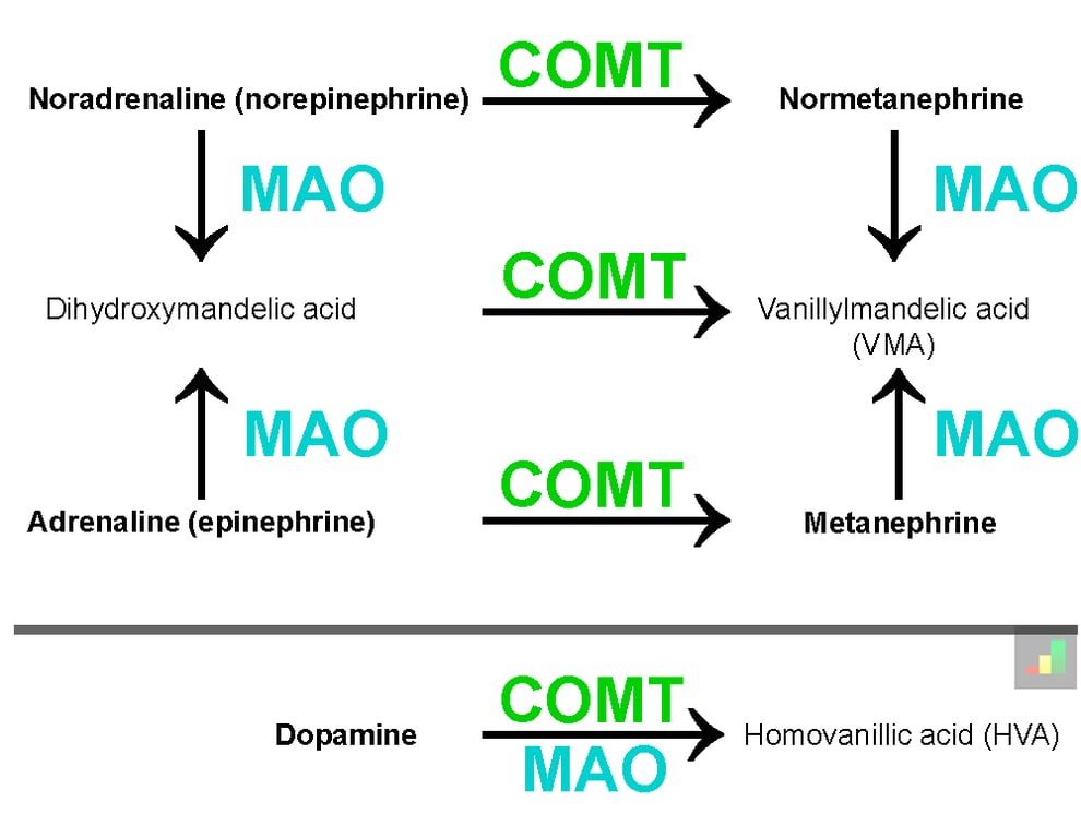 | |
Adrenaline and noradrenaline degradation pathways. Click on image to enlarge