Phaeochromocytoma
On this page
Definition
Phaeochromocytoma is a rare neuroendocrine tumour that secretes catecholamines. Phaeochromocytomas typically arise from chromaffin tissue in the adrenal medulla but in some cases arise in extra-adrenal chromaffin tissue (extra-adrenal phaeochromocytomas are sometimes termed paraganglioma).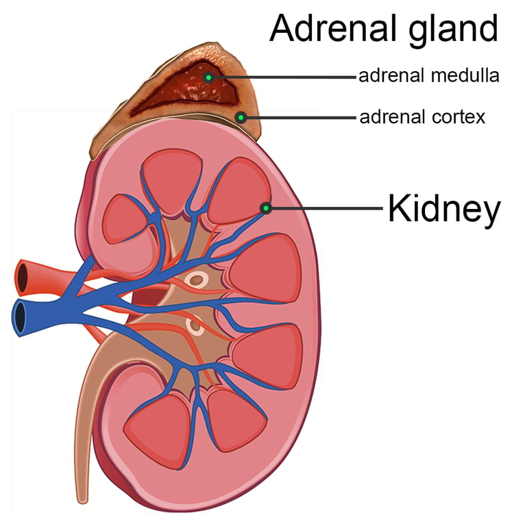 | |
Phaeochromocytomas typically arise from chromaffin tissue in the adrenal medulla. Click on image to enlarge
Background
The majority of phaeochromocytoma's arise spontaneously and typically present in those aged 40-50 years of age.
Up to 30% of symptomatic phaeochromocytoma and paraganglioma are hereditary and arise due to genetic mutations associated with the following conditions:
- MEN type 2
- Von Hippel-Lindau (VHL) disease
- Neurofibromatosis type 1
- Paraganglioma syndromes types 1, 3 and 4.
About 10% of all phaeochromocytoma and paraganglioma are metastatic. Extra-adrenal phaeochromocytoma (paragangliom) are more likely to be metatstatic.
Phaeochromocytoma & the 10% rule
It used to be quoted that:- 10% are extra-adrenal
- 10% are bilateral
- 10% are malignant
- 10% are found in children
- 10% are familial
- 10% are not associated with hypertension
Recent studies however suggest some of these figures are inaccurate, specifically:
- Up to 30% of cases are familial
- Up to 25% are malignant
- 15% of cases are extra-adrenal (most common site is the organ of Zuckerkandl, adjacent to the bifurcation of the aorta)
Clinical features
The clinical features of phaeochromocytoma are the result of excessive catecholamine production. Catecholamines include epinephrine (adrenaline), norepinephrine (noradrenaline), and dopamine.
The signs and symptoms are typically paroxysmal and include:
- Hypertension (around 90% of cases, may be sustained)
- Headaches
- Palpitations (tachycardia & cardiac arrhythmias)
- Sweating
- Anxiety (may describe fear or sense of impending doom)
- Tremor
- Abdominal & flank pain
- Nausea
Metabolic effects of catecholamines(caused by agonist activity at alpha and beta adrenoreceptors) |
|---|
| Glycogenolysis in liver and skeletal muscle Mobilisation of free fatty acids Increased serum lactate Increase in metabolic rate Increased myocardial force and rate of contraction Decrease in SVR (although vasoconstrictors epinephrine dilates vessels in skeletal muscle & liver leading to overall decrease in SVR) Increase in alertness and evokes feelings of anxiety and fear Rise in plasma glucose |
Investigations
- Plasma and urine metanephrines, catecholamines and urine vanillylmandelic acid (VMA)
- Abdominal CT or MRI often used to determine site
- If CT or MRI fail to find site a scan with metaiodobenzylguanidine (MIBG) labelled with radioactive iodine is performed (iodine 123 or 131). MIBG concentrates within adrenal tissue.
Blood test abnormalities associated with phaeochromocytoma include:
- Hyperglycaemia (common)
- Hypercalcaemia
- Polycythaemia (rare)
Plasma metanephrine assay (99% sensitivity with a specificity of 89%) has the highest sensitivity and specificity for phaeochromocytoma and is the diagnostic blood test of choice.
24 hr urinary collection of metanephrines has higher sensitivity (sensitivity 97%) than
24 hr urinary collection of catecholamines (sensitivity 86%) and has now largely replaced the later as the urinary test of choice.
24 hr urinary collection of metanephrines has higher sensitivity (sensitivity 97%) than
24 hr urinary collection of catecholamines (sensitivity 86%) and has now largely replaced the later as the urinary test of choice.
Management
Surgery is the definitive management. The patient must first however be stabilised with medical management:- Alpha-blocker e.g. phenoxybenzamine or phentolamine is given before beta blockade
- Beta-blocker e.g. propranolol
Alpha blockade with phenoxybenzamine is started at least 7 to 10 days before operation to allow for expansion of blood volume. Only once alpha blockade is achieved is beta blockade considered. If beta blockade is started too soon, unopposed alpha stimulation can precipitate a hypertensive crisis.