Distal convoluted tubule & collecting duct
On this page
The distal convoluted tubule (DCT) receives tubular fluid from the thick ascending limb of the loop of Henle. This fluid is hypotonic with an osmolality of ~ 90 mosmol/kg H2O when it enters the DCT.
The primary role of the DCT is regulation of potassium, sodium, calcium, and pH. The macula densa are also situated in the first segment of the DCT and play an important role in autoregulation.
Sodium reabsorption
The proximal convoluted tubule (PCT) and loop of Henle will have reabsorbed around 90% of filtered Na+ ions by the time the tubular fluid reaches the DCT. The remaining 10% is typically reabsorbed in the DCT and collecting duct under the influence of a number of hormones.
There are two main routes for Na+ reabsorption from the lumen:
Na+ ions are actively pumped out of the tubular cell cytosol into the interstitial fluid by the Na+-K+-ATPase on the basolateral membrane. This lowers the Na+ concentration within the cell and helps create the gradient for Na+ ions to move in via the above methods. Of note only NCC transporters are present in the early part of the DCT whereas both NCC and ENaC are present in the distal part of the DCT.
The movement of Na+ ions from the lumen into the cells is electrically offset by the excretion of K+ ions from cells into the lumen via renal potassium secretory channel ROMK (Renal outer medullary K+ channel) channels.
K+ is actively transported from the interstitial space into principal cells by basolateral Na+-K+-ATPase pumps then passively secreted via ROMK channels and K+/Cl- cotransporter. The relative negative luminal charge caused by Na+ reabsorption helps drive passive movement of K+ via ROMK channels. Secretion is driven by the concentration gradient between the cell cytosol and tubular fluid.
Secreted K+ is 'washed away' by tubular flow which helps maintain the concentration gradient between the lumen and cell cytosol for secretion. Increased tubular flow therefore increases potassium secretion.
ADH stimulates the insertion of aquaporin channels into the apical (lumen side) membrane. When ADH is present water diffuses from the lumen into principal cells through aquaporin channels and then diffuses across the basolateral membrane into the interstitial space. The loss of water concentrates the tubular fluid (which ultimately becomes urine) which reaches a maximum osmolality of 290 mOsmol/kg H2O by the end of the the cortical portion of the collecting duct.
The tubular fluid at this stage is radically different to plasma and the initial glomerular filtrate collected in Bowmans capsule as large quantities of Na+, K+, Cl- and HCO3- have been reabsorbed. The DCT and cortical portion of the collecting duct are impermeable to urea which concentrates as water is reabsorbed.
In the medullary portion of the collecting duct water is reabsorbed, in the presence of ADH, due to the high osmolality of the medullary interstitium. Note that the collecting ducts of all nephrons, both cortical and medullary, pass through the medulla and take part in concentrating urine.
In the medullary collecting duct urea diffuses along its concentration gradient into the medulla and back into the ascending loop of Henle. Urea is effectively trapped and cycles through the ascending loop, DCT and collecting duct again and again. Urea is a major contributor to the high osmolality of the medulla.
ADH increases medullary collecting duct permeability via activation of epithelial uniporters. This promotes the facilitated diffusion of urea out of the collecting duct and further concentrates urine by increasing medullary osmolality.
PTH activates Ca2+ channels in the apical (luminal) membrane that facilitate the movement of Ca2+ from the lumen into the cellular cytosol.
Ca2+ is transported from the cytosol into the interstitial fluid by two pumps in the basolateral membrane:
The Ca2+-ATPase pump on the basolateral membrane is stimulated by both PTH and activated vitamin D. In the cell binding proteins prevent free calcium ions from building up to dangerous levels.
The primary role of the DCT is regulation of potassium, sodium, calcium, and pH. The macula densa are also situated in the first segment of the DCT and play an important role in autoregulation.
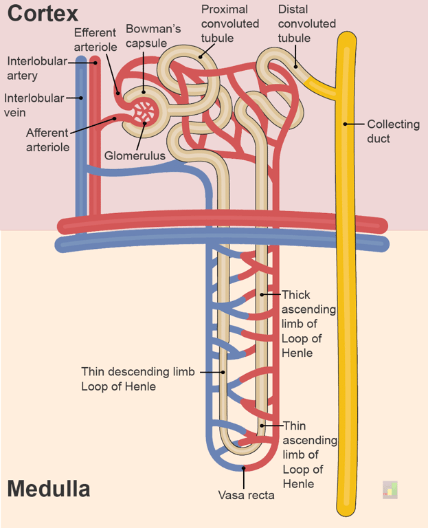 | |
The nephron. click on image to enlarge
Sodium reabsorption
The proximal convoluted tubule (PCT) and loop of Henle will have reabsorbed around 90% of filtered Na+ ions by the time the tubular fluid reaches the DCT. The remaining 10% is typically reabsorbed in the DCT and collecting duct under the influence of a number of hormones.
There are two main routes for Na+ reabsorption from the lumen:
- Na+ epithelial sodium channels (ENaC)
- NCC (sodium-chloride cotransporter)
Na+ ions are actively pumped out of the tubular cell cytosol into the interstitial fluid by the Na+-K+-ATPase on the basolateral membrane. This lowers the Na+ concentration within the cell and helps create the gradient for Na+ ions to move in via the above methods. Of note only NCC transporters are present in the early part of the DCT whereas both NCC and ENaC are present in the distal part of the DCT.
ENaC channels are inhibited by atrial natriuretic peptide (ANP)
The movement of Na+ ions from the lumen into the cells is electrically offset by the excretion of K+ ions from cells into the lumen via renal potassium secretory channel ROMK (Renal outer medullary K+ channel) channels.
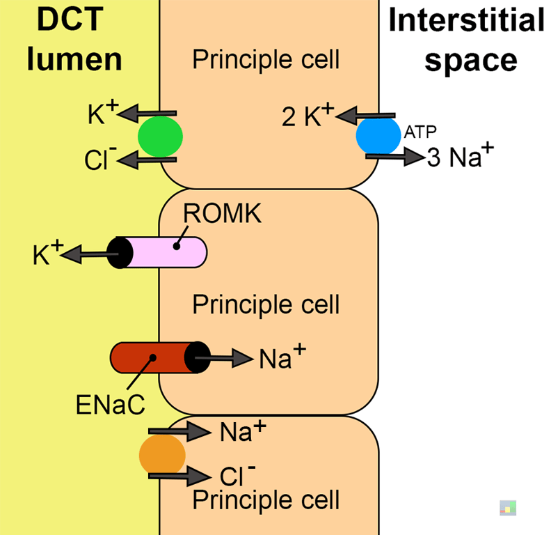 | |
Ion transport in the principle cells of the DCT. Click on image to enlarge
Aldosterone and the DCT
Aldosterone has a number of effects on the DCT cells:
The overall effect is to drive Na+ ions from the lumen into the interstitial fluid and drive K+ ions in the opposite direction.
Aldosterone has a number of effects on the DCT cells:
- Increases activity of the Na+-K+-ATPase on the basolateral membrane uptake into the principle cells
- Increases expression of ROMK channels
- Increases expression of ENaC channels
The overall effect is to drive Na+ ions from the lumen into the interstitial fluid and drive K+ ions in the opposite direction.
Potassium secretion
Reabsorption of potassium is typically complete by the time the tubular fluid reaches the DCT. Potassium secretion is primarily regulated by the cells of the late distal tubule. As can be seen from the notes above K+ secretion is linked to Na+ reabsorption.K+ is actively transported from the interstitial space into principal cells by basolateral Na+-K+-ATPase pumps then passively secreted via ROMK channels and K+/Cl- cotransporter. The relative negative luminal charge caused by Na+ reabsorption helps drive passive movement of K+ via ROMK channels. Secretion is driven by the concentration gradient between the cell cytosol and tubular fluid.
Secreted K+ is 'washed away' by tubular flow which helps maintain the concentration gradient between the lumen and cell cytosol for secretion. Increased tubular flow therefore increases potassium secretion.
Diuretics that increase tubular flow increase K+ loss and may result in hypokalaemia
Potassium & acid-base homeostasis
In general, acidosis causes decreased K+ secretion and increased reabsorption in the collecting duct. Alkalosis has the opposite effects, often leading to hypokalemia. Potassium disorders also influence acid-base homeostasis. Potassium depletion causes increased H+ secretion, ammoniagenesis and H-K-ATPase activity.Water reabsorption
The cells of the DCT and collecting duct are impermeable to water except in the presence of antidiuretic hormone (ADH).ADH stimulates the insertion of aquaporin channels into the apical (lumen side) membrane. When ADH is present water diffuses from the lumen into principal cells through aquaporin channels and then diffuses across the basolateral membrane into the interstitial space. The loss of water concentrates the tubular fluid (which ultimately becomes urine) which reaches a maximum osmolality of 290 mOsmol/kg H2O by the end of the the cortical portion of the collecting duct.
The tubular fluid at this stage is radically different to plasma and the initial glomerular filtrate collected in Bowmans capsule as large quantities of Na+, K+, Cl- and HCO3- have been reabsorbed. The DCT and cortical portion of the collecting duct are impermeable to urea which concentrates as water is reabsorbed.
In the medullary portion of the collecting duct water is reabsorbed, in the presence of ADH, due to the high osmolality of the medullary interstitium. Note that the collecting ducts of all nephrons, both cortical and medullary, pass through the medulla and take part in concentrating urine.
The final urine osmolality may reach concentrations of 1400 mOsmol/kg H2O under maximal ADH stimulation.
In the absence of ADH urine concentrations may be dilute with concentrations as low as 60 mOsmol/kg H2O. This may occur pathologically in cases of
In the absence of ADH urine concentrations may be dilute with concentrations as low as 60 mOsmol/kg H2O. This may occur pathologically in cases of
diabetes insipidus
Urea
Whilst the DCT and cortical portion of the collecting duct are impermeable to urea, the medullary portion of the collecting duct is relatively permeable to urea.In the medullary collecting duct urea diffuses along its concentration gradient into the medulla and back into the ascending loop of Henle. Urea is effectively trapped and cycles through the ascending loop, DCT and collecting duct again and again. Urea is a major contributor to the high osmolality of the medulla.
Urea provides ~50% of osmolality in the renal medulla
ADH increases medullary collecting duct permeability via activation of epithelial uniporters. This promotes the facilitated diffusion of urea out of the collecting duct and further concentrates urine by increasing medullary osmolality.
Calcium reabsorption
Calcium reabsorption in the DCT is regulated by parathyroid hormone (PTH) and 1,25 dihydroxycholecalciferol (activated vitamin D).PTH activates Ca2+ channels in the apical (luminal) membrane that facilitate the movement of Ca2+ from the lumen into the cellular cytosol.
Ca2+ is transported from the cytosol into the interstitial fluid by two pumps in the basolateral membrane:
- Ca2+-ATPase pump
- Na+-Ca2+ antiporter
The Ca2+-ATPase pump on the basolateral membrane is stimulated by both PTH and activated vitamin D. In the cell binding proteins prevent free calcium ions from building up to dangerous levels.
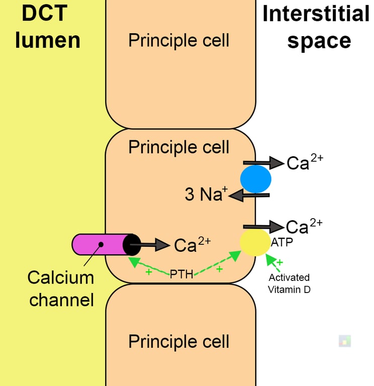 | |
Calcium ion reabsorption in the distal convoluted tubule. Click on image to enlarge
PTH inhibits phosphate reabsorption.
This is clinically relevant as conditions such as primary hyperparathyroidism will cause low phosphate.
This is clinically relevant as conditions such as primary hyperparathyroidism will cause low phosphate.