Renal circulation
The kidneys play a key role in filtering blood and modifying its composition via reabsorption and secretion.
To perform this role they are highly vascularised and receive a high percentage of cardiac output..
The renal arteries enter each kidney at the hilus. The renal arteries first divide into segmental arteries, followed by further branching into interlobar arteries that pass through the renal columns to reach the cortex. The interlobar arteries terminate by branching into arcuate arteries. The arcuate arteries give off branches called interlobular arteries which are also known as cortical radiate arteries. The interlobular arteries ascend the cortex and branch into the afferent arterioles.
The afferent arterioles form part of the glomerulus. They feed into the glomerular capillary network which is the site of filtration. Following filtration blood drains from the capillary network into efferent arterioles rather than venules as this allows greater hydrostatic pressure to be generated within the glomerulus to provide the force for ultrafiltration. Afferent and efferent arterioles provide the major resistance to renal blood flow.
Efferent arterioles then further branch into capillary networks, called peritubular capillaries, in the cortex around the proximal and distal tubules. Capillaries close to the boundary between the renal cortex and medulla loop into the medulla forming vasa recta which surround the loop of Henle. The vasa recta drain into venules which ultimately drain back to the renal vein following the same pattern as the renal arteries but in reverse (see the image above).
Renal blood flow & autoregulation
The kidneys receive approximately 20% of cardiac output. The renal cortex receives around 90% of renal blood flow (~ 500 mL/min/100g) with the medulla receiving the remainder (20-100 mL/min/100g).
Renal blood flow is subject to a high degree of autoregulation which is achieved via differential constriction of afferent and efferent arterioles through the myogenic response and mediated by the macula densa.
The myogenic mechanism or response is one method of autoregulation where high pressure within the renal arterioles stretches arteriole smooth muscle and stimulates a reflex vasoconstriction to reduce renal blood flow.
The macula densa are specialised cells at the junction of the thick ascending loop of Henle and the proximal part of the distal convoluted tubule. The macula densa take part in the juxtaglomerular apparatus. They are chemoreceptors that detect the concentration of sodium chloride. A decrease in sodium chloride concentration indicates low plasma flow and the macula densa respond by:
Hormonal and chemical mediators affecting renal blood flow
Note that autoregulation refers to maintaining a constant renal blood flow over a range of blood pressures. A number of other hormones may have effects on renal blood flow.
Several hormones and chemical mediators promote vasoconstriction and therefore decrease renal blood flow. These include:
A number of hormones and chemical mediators lower arterial resistance via vasodilation and increase renal blood flow. These include:
To perform this role they are highly vascularised and receive a high percentage of cardiac output..
The kidneys receive approximately 20% of cardiac output.
90% of blood entering the kidneys supplies the cortex.
90% of blood entering the kidneys supplies the cortex.
Arterial supply
The renal arteries are paired branches of the aorta that arise from the left interior side of the abdominal aorta, immediately below the superior mesenteric artery.The renal arteries enter each kidney at the hilus. The renal arteries first divide into segmental arteries, followed by further branching into interlobar arteries that pass through the renal columns to reach the cortex. The interlobar arteries terminate by branching into arcuate arteries. The arcuate arteries give off branches called interlobular arteries which are also known as cortical radiate arteries. The interlobular arteries ascend the cortex and branch into the afferent arterioles.
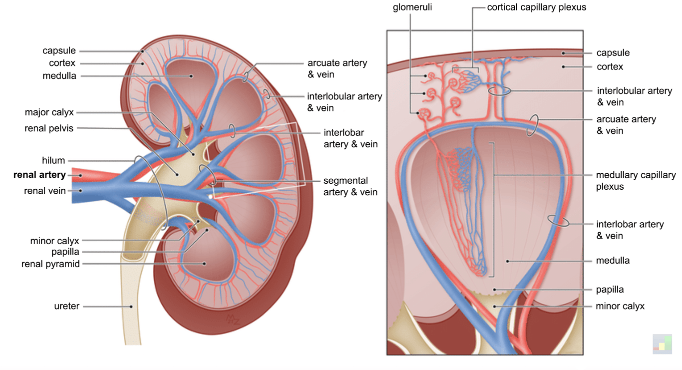 | |
Arterial supply of the kidney. click on image to enlarge
The afferent arterioles form part of the glomerulus. They feed into the glomerular capillary network which is the site of filtration. Following filtration blood drains from the capillary network into efferent arterioles rather than venules as this allows greater hydrostatic pressure to be generated within the glomerulus to provide the force for ultrafiltration. Afferent and efferent arterioles provide the major resistance to renal blood flow.
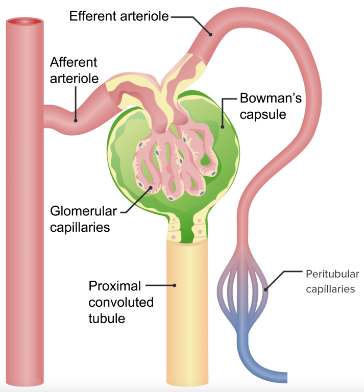 | |
Afferent and efferent arterioles arrangement in the kidney nephron. click on image to enlarge
Efferent arterioles then further branch into capillary networks, called peritubular capillaries, in the cortex around the proximal and distal tubules. Capillaries close to the boundary between the renal cortex and medulla loop into the medulla forming vasa recta which surround the loop of Henle. The vasa recta drain into venules which ultimately drain back to the renal vein following the same pattern as the renal arteries but in reverse (see the image above).
The vasa recta are the only blood supply to the renal medulla
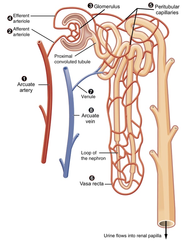 | |
Afferent and efferent arterioles and the vasa recta of the nephron. click on image to enlarge
Renal blood flow & autoregulation
The kidneys receive approximately 20% of cardiac output. The renal cortex receives around 90% of renal blood flow (~ 500 mL/min/100g) with the medulla receiving the remainder (20-100 mL/min/100g).
Renal blood flow is subject to a high degree of autoregulation which is achieved via differential constriction of afferent and efferent arterioles through the myogenic response and mediated by the macula densa.
The myogenic mechanism or response is one method of autoregulation where high pressure within the renal arterioles stretches arteriole smooth muscle and stimulates a reflex vasoconstriction to reduce renal blood flow.
The macula densa are specialised cells at the junction of the thick ascending loop of Henle and the proximal part of the distal convoluted tubule. The macula densa take part in the juxtaglomerular apparatus. They are chemoreceptors that detect the concentration of sodium chloride. A decrease in sodium chloride concentration indicates low plasma flow and the macula densa respond by:
- Dilating afferent arterioles, raising glomerular hydrostatic pressure and increasing blood flow
- Increases renin release from the juxtaglomerular cells of the afferent and efferent arterioles which promotes vasoconstriction.
- Releasing adenosine which diffuses across to afferent arterioles and increases vascular resistance via vasoconstriction
The juxtaglomerular apparatus consists of three types of cells:
- Macula densa - part of the thick ascending limb of the loop of Henle an the proximal distal convoluted tubule of the same nephron
- Juxtaglomerular cells (aka granular cells) - specialised cells mainly found in the walls of afferent arterioles that secrete renin
- Extraglomerular mesangial cells - function of these cells not fully understood but they contain smooth muscle and are thought to play a role in support and regulate renal blood flow.
Hormonal and chemical mediators affecting renal blood flow
Note that autoregulation refers to maintaining a constant renal blood flow over a range of blood pressures. A number of other hormones may have effects on renal blood flow.
Several hormones and chemical mediators promote vasoconstriction and therefore decrease renal blood flow. These include:
- Noradrenaline (norepinephrine) constricts both afferent and efferent arterioles
- Adrenaline (epinephrine) constricts both afferent and efferent arterioles
- Angiotensin II (end product of renin synthesis) constricts both afferent and efferent arterioles but efferent >afferent
- Adenosine
- Endothelin
A number of hormones and chemical mediators lower arterial resistance via vasodilation and increase renal blood flow. These include:
- Atrial natriuretic peptide
- Brain natriuretic peptide
- Prostacyclin (also called prostaglandin I2 or PGI2)
- Bradykinin
- Dopamine (may cause vasoconstriction at high doses)