Macroscopic structure of the kidney
On this page
Position
The kidney's are retroperitoneal organs located on either side of the vertebral column and typically extend from T12- L3. The right kidney often sits slightly lower than the left due to the liver. Each kidney typically spans three vertebrae in height.Structure
Each kidney is covered in a tough, fibrous outer layer of connective tissue called the capsule that helps protect the kidney's soft inner tissues. Deep to the capsule there is an outer, lighter-colored region called the renal cortex and an inner darker-colored region called the medulla. The cortex and medulla are the 2 main functional sections of the kidney. The third section is composed of calyces and renal pelvis which collect urine and are continuous with the ureter.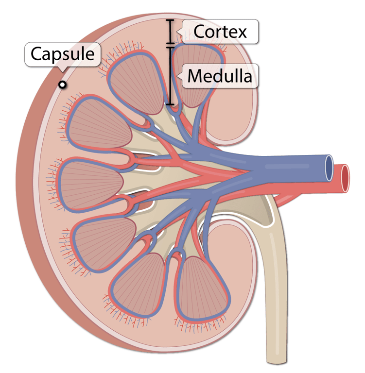 | |
Image showing renal cortex and medulla. Click on image to enlarge
Cortex and medulla
The renal cortex contains the glomeruli and is the site of filtration. The proximal and distal convoluted tubules are also contained within the cortex. The renal medulla contains the collecting ducts, loops of Henle, vasa recta and medullary interstitium. The renal medulla is split up into a number of sections, known as the renal pyramids (more below).
Contents of the cortex and medulla
| Cortex | Medulla |
|---|---|
| Glomeruli Bowman's capsule Proximal convoluted tubule Distal convoluted tubule | Loop of Henle Collecting ducts Vasa recta Medullary interstitium |
Lobes, pyramids and columns
Each kidney contains approximately 6-8 renal lobes, with each lobe containing thousands of nephrons (the nephron is the kidney's functional unit).
The medullary portion of a renal lobe is called a renal pyramid due to its shape. The base (wide end) of a pyramid lies adjacent to the renal cortex, and the tip or papilla (narrow end) extends into the kidney's medial indentation. A pyramid appears darker and somewhat striated because it contains a high concentration of straight, parallel tubules called collecting ducts, which transport urine to a lobe's apex.
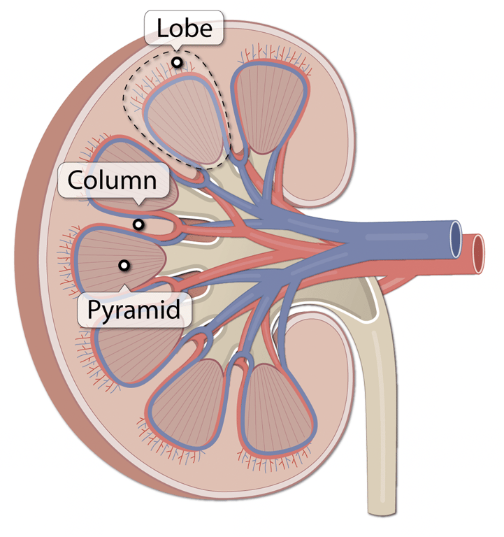 | |
Image showing renal cortex and medulla. Click on image to enlarge
A renal column extends between and separates adjacent renal lobes. The columns largely consist of connective tissue and provide a supportive framework for blood vessels that enter and exit the cortex.
Calyces, renal pelvis and ureters
The papilla of each pyramid empties urine into a minor calyx. Minor calyces empty into major calyces, and major calyces empty into the renal pelvis. This becomes the ureter. Urine is propelled through the ureter into the bladder by peristalsis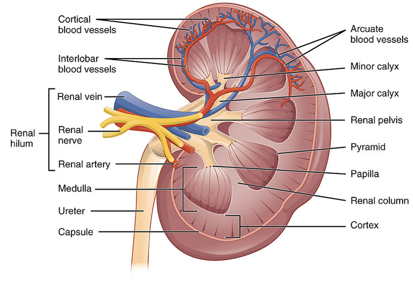 | |
Image showing overall anatomy of the kidney. Click on image to enlarge