Haemoglobin
On this page
Haemoglobin
Haemoglobin synthesis requires iron, folate and vitamin B12.Each haemoglobin molecule contains 4 protein chains, sometimes referred to as globin subunits. Each protein chain contains a ring-like heme group containing an iron atom.
Haemoglobin variants exist with differences in the globin subunits. These variants are a normal part of embryonic and fetal development but pathological variants also exist.
Normal adult haemoglobin contains two alpha and two beta chains.
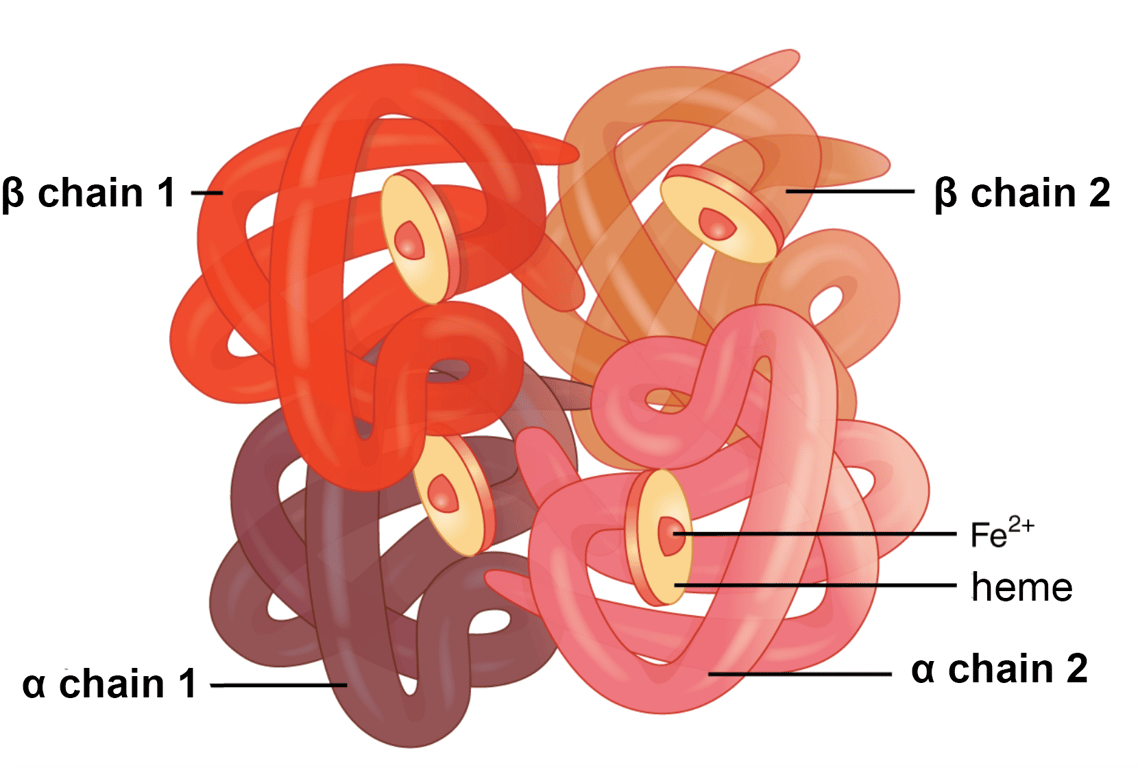 | |
Normal adult haemoglobin structure. click on image to enlarge
Embryonic and fetal haemoglobin
| Embryonic Haemoglobin | Fetal Haemoglobin |
|---|---|
|
Adult haemoglobin
The main adult haemoglobin (HbA or HbA1) is composed of two alpha & two beta globin subunits. It is sometimes written as α2β2. HbA1 typically accounts for 97% or more of haemoglobin in most adults.
HbA2 is composed of two alpha and two delta globin subunits and may be written as α2Δ2. HbA2 makes up 1-3% of haemoglobin in an adult. A small amount of HbF is often present in adults and typically accounts for 0.5-2% of the total.
Red blood cell & haemoglobin destruction
Red blood cells typically circulate for around 120 days before they are destroyed by macrophages in the liver and spleen.The haem (or heme) group is converted into bilverdin then bilirubin. Iron is recycled via the transport protein transferrin or stored as ferritin. Increased red cell destruction may lead to elevated bilirubin levels and jaundice.