Gastrointestinal physiology: Functional anatomy of the GI tract
On this page
The wall of the gastrointestinal tract (GIT) consists of four layers of specialised tissue. From inside (lumen) to out these are:
Mucosa
The mucosa is the innermost layer of the GIT. The mucosa is divided into three sublayers:
The epithelium is the innermost layer which comes into contact with chyme. Here is where digestive, absorptive and secretory processes occur. The lamina propria is a layer of connective tissue whilst the muscularis mucosae is a thin layer of smooth muscle
The epithelium contains a high number of mucus secreting goblet cells which aids the passage of food through the GIT and forms a protective barrier. The epithelium may contain adaptations depending on the which part of the GIT it is. In the small intestine for example the epithelium is folded into villi and contains projections called microvilli to maximise surface area for absorption. In the large intestine villi are absent.
Underlying the epithelium is the lamina propria, which contains myofibroblasts, blood vessels, lymph vessels, nerves, and several different immune cells.
Muscularis mucosa is a thin layer of smooth muscle that aids in the action of continued peristalsis and catastalsis along the gut.
Between the two muscle layers is the myenteric nerve plexus (also known as Auerbach's plexus). The myenteric plexus sends impulses to the muscle layers which result in peristalsis. Activity is initiated by the pacemaker cells (interstitial cells of Cajal). The gut has intrinsic peristaltic activity but this may be modulated by the input from the autonomic nervous system.
The coordinated contractions of these layers results in peristalsis which propels food/chyme/faeces through the GIT. Food in the GI tract is called a bolus (ball of food) from the mouth down to the stomach. The circular muscle layer contracts behind any food bolus /chyme/faeces which prevents food travelling backwards.
In different parts of the GIT the muscle layer is adapted e.g. the muscular layer of the colon is much thicker to handle the more viscous heavy faeces. In fact in the colon the longitudinal layers of muscle form 3 thickened bands called taeniae coli which result in haustration.
Serosa
The outermost layer of the GIT wall consists of several layers of connective tissue called adventitia or serosa.
Regions of the gastrointestinal tract within the peritoneum (called intraperitoneal) are covered with serosa. This structure consists of connective tissue covered by a simple squamous epithelium, called the mesothelium, which reduces frictional forces during digestive movements. The intraperitoneal regions include most of the stomach, first part of the duodenum, all of the small intestine, caecum and appendix, transverse colon, sigmoid colon and rectum. In these sections of the gut there is clear boundary between the gut and the surrounding tissue. These parts of the tract have a mesentery.
Regions of the GIT behind the peritoneum (called retroperitoneal) are covered with adventitia. They blend into the surrounding tissue and are fixed in position (for example, the retroperitoneal section of the duodenum usually passes through the transpyloric plane). The retroperitoneal regions include the oral cavity, oesophagus, pylorus of the stomach, distal duodenum, ascending colon, descending colon and anal canal
- Mucosa
- Submucosa
- Muscular layer
- Serosa (adventitia)
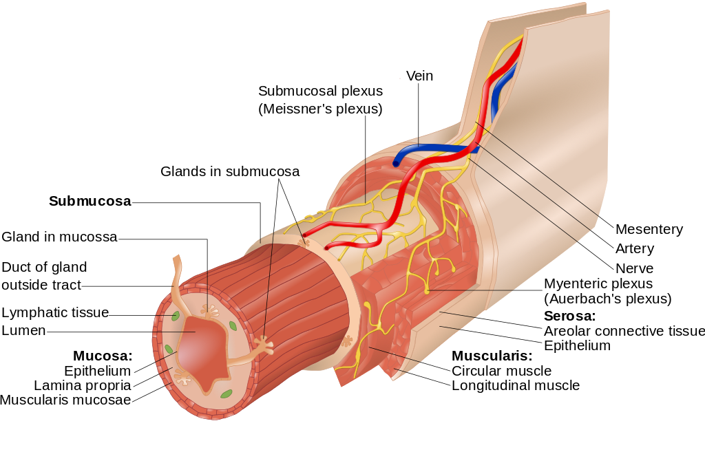 | |
Illustration of layers of the GI tract. click on image to enlarge
Mucosa
The mucosa is the innermost layer of the GIT. The mucosa is divided into three sublayers:
- Epithelium (innermost)
- Lamina propria (connective tissue)
- Muscularis mucosae (smooth muscle layer)
The epithelium is the innermost layer which comes into contact with chyme. Here is where digestive, absorptive and secretory processes occur. The lamina propria is a layer of connective tissue whilst the muscularis mucosae is a thin layer of smooth muscle
The epithelium contains a high number of mucus secreting goblet cells which aids the passage of food through the GIT and forms a protective barrier. The epithelium may contain adaptations depending on the which part of the GIT it is. In the small intestine for example the epithelium is folded into villi and contains projections called microvilli to maximise surface area for absorption. In the large intestine villi are absent.
Underlying the epithelium is the lamina propria, which contains myofibroblasts, blood vessels, lymph vessels, nerves, and several different immune cells.
Muscularis mucosa is a thin layer of smooth muscle that aids in the action of continued peristalsis and catastalsis along the gut.
Submucosa
The submucosa consists of a dense irregular layer of connective tissue with blood vessels, lymphatics, and nerves (submucosal or Meissner's plexus) whose branches penetrate into the mucosa and innervate the cells in the epithelial layer and the smooth muscle of the muscularis mucosae.The submucosa contains the submucous plexus and enteric nervous plexus situated on the inner surface of the muscular layer
Muscular layer
The muscular layer (also known as the muscularis propria and muscularis externa) consists of two layers of muscle. The inner layer is composed of circular muscle whilst the outer muscle layer is organised longitudinally (the stomach has an extra inner oblique muscle layer).Between the two muscle layers is the myenteric nerve plexus (also known as Auerbach's plexus). The myenteric plexus sends impulses to the muscle layers which result in peristalsis. Activity is initiated by the pacemaker cells (interstitial cells of Cajal). The gut has intrinsic peristaltic activity but this may be modulated by the input from the autonomic nervous system.
The coordinated contractions of these layers results in peristalsis which propels food/chyme/faeces through the GIT. Food in the GI tract is called a bolus (ball of food) from the mouth down to the stomach. The circular muscle layer contracts behind any food bolus /chyme/faeces which prevents food travelling backwards.
In different parts of the GIT the muscle layer is adapted e.g. the muscular layer of the colon is much thicker to handle the more viscous heavy faeces. In fact in the colon the longitudinal layers of muscle form 3 thickened bands called taeniae coli which result in haustration.
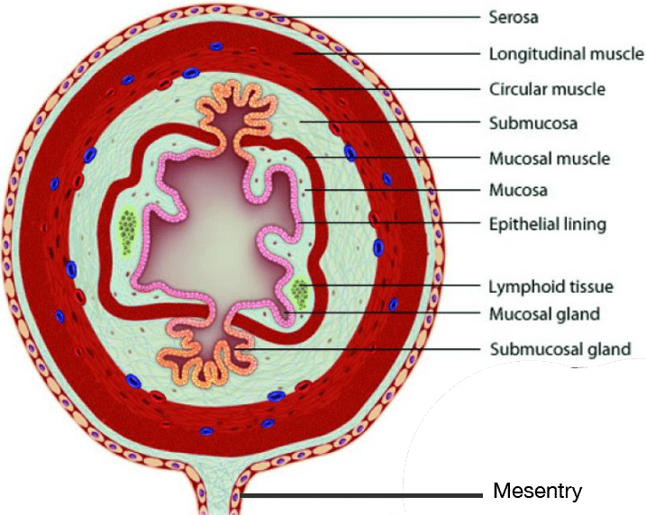 | |
Layers of the GI tract illustrated. Click on image to enlarge
Serosa
The outermost layer of the GIT wall consists of several layers of connective tissue called adventitia or serosa.
Regions of the gastrointestinal tract within the peritoneum (called intraperitoneal) are covered with serosa. This structure consists of connective tissue covered by a simple squamous epithelium, called the mesothelium, which reduces frictional forces during digestive movements. The intraperitoneal regions include most of the stomach, first part of the duodenum, all of the small intestine, caecum and appendix, transverse colon, sigmoid colon and rectum. In these sections of the gut there is clear boundary between the gut and the surrounding tissue. These parts of the tract have a mesentery.
Regions of the GIT behind the peritoneum (called retroperitoneal) are covered with adventitia. They blend into the surrounding tissue and are fixed in position (for example, the retroperitoneal section of the duodenum usually passes through the transpyloric plane). The retroperitoneal regions include the oral cavity, oesophagus, pylorus of the stomach, distal duodenum, ascending colon, descending colon and anal canal