Gastrointestinal physiology: Small intestine
On this page
The small intestines comprise the duodenum, jejunum and ileum. The primary function of the small intestines is digestion of food and absorption of the by products.
Chyme that enters the duodenum is acidic, hypertonic and only partly digested which prevents absorption of its breakdown products.
Functional anatomy of the intestinal villus
The small intestine has a number of adaptations to increase its surface area for absorption:
Other features that ensure the small intestine is highly efficient at its function include:
Each villus has a blind ended lymphatic channel called a lacteal and a capillary network between arterioles and venules. Nutrients are absorbed into the blood stream here. Blood from the small intestine ultimately passes into the portal circulation and into the liver where further catabolism occurs.
The sodium pump expels 3 Na+ ions from the cell into the lumen in exchange for 2 K+ ions with both ions moving against their concentration gradients.
The small intestine releases enzymes to facilitate the enzymatic breakdown of larger chain carbohydrates into these short chain sugars.
The brush border releases the following enzymes:
Once broken down into monosaccharides transport proteins facilitate their movement from the intestinal lumen across the apical membrane into the cells of the intestine (enterocytes). These transporters are either SGLT (sodium glucose co-transporters) or GLUT (glucose co-transporters) type. SGLT co-transporters primarily move glucose and galactose across the apical membrane and are an example of secondary active transport utilising the energy of moving Na+ ions along its concentration gradient to drive these monosaccharides in the same direction. Fructose is moved across the apical membrane by a GLUT transporter which utilises facilitated diffusion (a form of passive transport).
Monosaccharides exit the enterocytes to enter the bloodstream via facilitated diffusion across the basolateral (basal) membrane. This is a passive process via GLUT transporters (see image below).
The pancreatic enzymes trypsin and chemotrypsin are secreted into the duodenum and break down polypeptides into oligopeptides.
The pancreatic enzyme carboxypeptidase and luminal epithelial enzyme aminopeptidase then break oligopeptides into amino acids. This is the final stage of protein digestion and occurs on the small intestine brush border.
Amino acids are transported across the epithelial membrane via secondary active transport (co-transport) with Na+ ions (sodium moves along its concentration gradient which drives the transporter to move amino acids across the epithelial membrane against their concentration gradient). Amino acids cross the basal membrane of the enterocyte into the capillaries by facilitated diffusion.
Calcium handling
The vast majority of calcium in our bodies is stored in bones or bound to proteins. Free calcium levels are typically very low so free calcium that is ingested diffuses across the luminal membrane into epithelial cells through membrane channels or via transporter proteins along a steep concentration gradient. Once in the cell calcium ions will bind to proteins that transport them from the luminal membrane to the basolateral membrane where it is actively transported by one of the following two transporters:
Note an antiporter may also be referred to as an exchanger and involves two ions moving across the membrane in opposite directions. It often utilises secondary active transport i.e. one of the ions is moving along their concentration gradients and one is moved against theirs.
For absorption to occur iron forms a complex with ascorbate. The resulting complex is transported across the epithelial membrane by a carrier protein called divalent metal transporter 1 (DMT1). Once in the cell iron may bind to a number of substances such as ferritin. A second transport protein (ferroportin) transports iron across the basolateral membrane into the bloodstream. In the bloodstream iron binds with transferrin and most is transported to the bone marrow for erythropoiesis.
Macrophages of the reticuloendothelial system store iron as part of the process of breaking down haemoglobin from engulfed red blood cells. Iron may be stored in two forms:
Iron absorption is regulated by the hormone hepcidin which is synthesised by the liver. Hepcidin reduces iron absorption into the portal system in two ways:
Emulsified fat is then broken down into free fatty acids and monoglycerides (fatty acid + glycerol) by lipase. As can be seen from the diagram below bile acids are involved in multiple steps in the process of fat digestion. After lipase has broken the fat into fatty acids and monoglycerides it combines with bile acids to form micelles.
Micelles have a hydrophilic shell and a hydrophobic core which allows the micelle to enter the aqueous layer of the microvilli allowing the following molecules to diffuse passively into the duodenal cells:
The bile acids remain in the intestinal lumen and travel to the terminal ileum where they are reabsorbed.
The micelles facilitate the transport of fatty acids and monoglycerides from the lumen across the epithelial membrane and into the cell. Once inside the enterocyte, fatty acids and monoglyceride are transported into the endoplasmic reticulum, where they are used to synthesise triglyceride. Beginning in the endoplasmic reticulum and continuing in the Golgi, triglyceride is packaged with cholesterol, lipoproteins and other lipids into particles called chylomicrons. Chylomicrons exit the cell by exocytosis across the basolateral membrane, they are too large to diffuse through capillaries and instead enter the lymphatics via endothelial gaps in the lacteals. The lymph is returned to the venous circulation via the lymphatic duct.
Water soluble vitamins are typically absorbed via simple diffusion except vitamin B12 which is bound by intrinsic factor and absorbed in the terminal ileum via endocytosis.
Duodenum
Chyme passes from the stomach into the duodenum via the pylorus. This triggers a number of responses to inhibit gastric juice release and gastric emptying. These responses include the release of gastric inhibitory hormones such as secretin, gastric inhibitory peptide (GIP) and cholecystokinin (CCK) as well as vagal and local reflexes.Chyme that enters the duodenum is acidic, hypertonic and only partly digested which prevents absorption of its breakdown products.
As chyme transits through the duodenum there is an osmotic movement of water which transforms chyme into an isotonic solution. There is also addition of bicarbonate from bile and pancreatic juices to neutralise the acidity. Further digestion is via enzymes released from the pancreas, liver (gallbladder) and intestine.
Functional anatomy of the intestinal villus
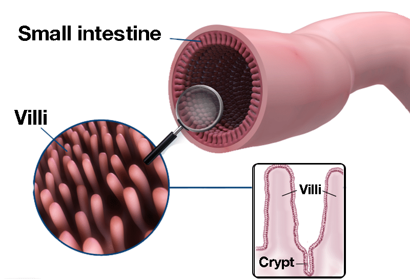 | |
Overview of small intestine structure at different magnifications. Note the presence of crypts between villi. Click on image to enlarge
The small intestine has a number of adaptations to increase its surface area for absorption:
- Mucosa is highly folded (plicae circulares)
- Contains finger like projections called villi
- Long length of small intestine for absorption (approx 4m)
- Presence of multiple small extensions to cells called microvilli form the brush border
Other features that ensure the small intestine is highly efficient at its function include:
- Crypts secrete 3L of hypotonic fluid per day
- Chyme is forced to flow in circular motion promoting mixing and contact with epithelial cells
- High turnover of epithelial cells with cells turning over every 5-6 days
- Thin epithelial membrane
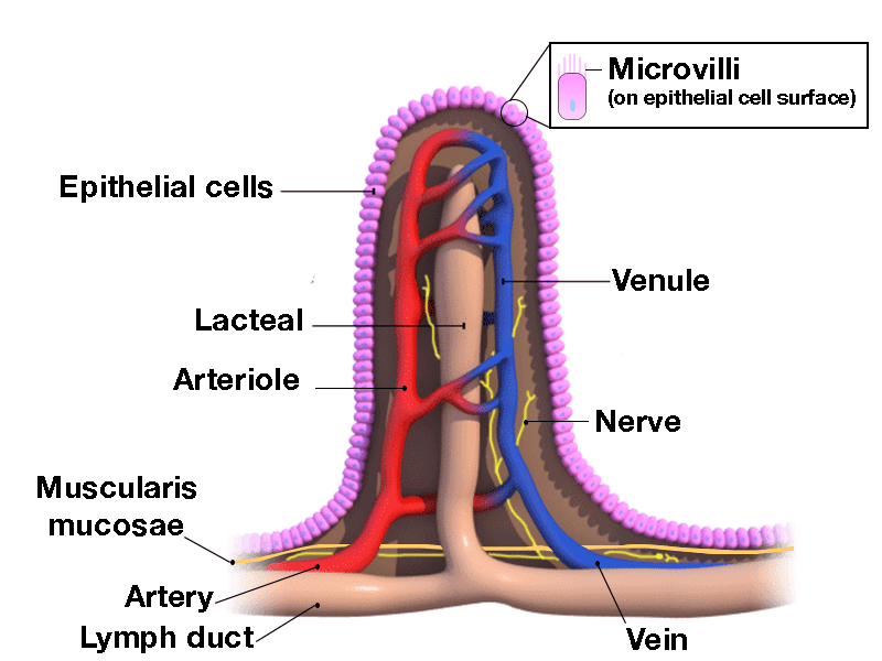 | |
Structure of villi within small intestine. Note the microvilli present on the magnification (brush border). Click on image to enlarge
Each villus has a blind ended lymphatic channel called a lacteal and a capillary network between arterioles and venules. Nutrients are absorbed into the blood stream here. Blood from the small intestine ultimately passes into the portal circulation and into the liver where further catabolism occurs.
The sodium pump
As chyme becomes isotonic there is no natural osmotic gradient between the luminal fluid and the cells of the small intestine. An osmotic gradient must therefore be created by the active movement of ions. This is achieved by the sodium pump- The sodium pump is an active Na+-K+-ATPase transporter in the small intestine.
The sodium pump expels 3 Na+ ions from the cell into the lumen in exchange for 2 K+ ions with both ions moving against their concentration gradients.
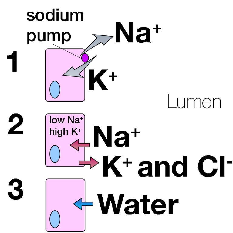 | |
1. The sodium pump actively pumps sodium ions out of the cell against its concentration gradient in exchange for potassium.
2. This leaves the cell low in sodium ions and high in potassium ions. These ions then start to flow along the newly created concentration gradients through membrane channels and via transport protein mechanisms. Note chloride moves out of the cell along with potassium.
3. Water follows the flow of sodium ions into the cell
Carbohydrate handling
Carbohydrates are absorbed as monosaccharides i.e. glucose, fructose and galactose.The small intestine releases enzymes to facilitate the enzymatic breakdown of larger chain carbohydrates into these short chain sugars.
The brush border releases the following enzymes:
- Maltases
- Isomaltases
- Sucrase
- Lactase
Once broken down into monosaccharides transport proteins facilitate their movement from the intestinal lumen across the apical membrane into the cells of the intestine (enterocytes). These transporters are either SGLT (sodium glucose co-transporters) or GLUT (glucose co-transporters) type. SGLT co-transporters primarily move glucose and galactose across the apical membrane and are an example of secondary active transport utilising the energy of moving Na+ ions along its concentration gradient to drive these monosaccharides in the same direction. Fructose is moved across the apical membrane by a GLUT transporter which utilises facilitated diffusion (a form of passive transport).
Monosaccharides exit the enterocytes to enter the bloodstream via facilitated diffusion across the basolateral (basal) membrane. This is a passive process via GLUT transporters (see image below).
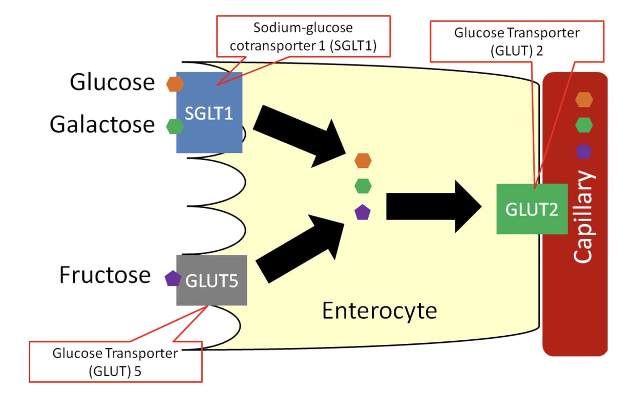 | |
Carbohydrate absorption in the intestine. Click on image to enlarge
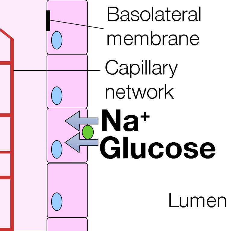 | |
Diagram illustrating sodium-monosaccharide co-transporter (SGLT). Green ball represents the co-transporter. Click to enlarge
Protein handling
The digestion of dietary protein starts in the stomach where protein is broken down into polypeptides by pepsin.The pancreatic enzymes trypsin and chemotrypsin are secreted into the duodenum and break down polypeptides into oligopeptides.
The pancreatic enzyme carboxypeptidase and luminal epithelial enzyme aminopeptidase then break oligopeptides into amino acids. This is the final stage of protein digestion and occurs on the small intestine brush border.
Amino acids are transported across the epithelial membrane via secondary active transport (co-transport) with Na+ ions (sodium moves along its concentration gradient which drives the transporter to move amino acids across the epithelial membrane against their concentration gradient). Amino acids cross the basal membrane of the enterocyte into the capillaries by facilitated diffusion.
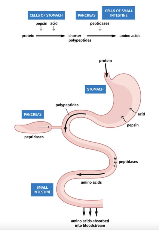 | |
Summary of protein digestion. Click on image to enlarge
Calcium handling
The vast majority of calcium in our bodies is stored in bones or bound to proteins. Free calcium levels are typically very low so free calcium that is ingested diffuses across the luminal membrane into epithelial cells through membrane channels or via transporter proteins along a steep concentration gradient. Once in the cell calcium ions will bind to proteins that transport them from the luminal membrane to the basolateral membrane where it is actively transported by one of the following two transporters:
- Ca2+ - ATPase active transporter
- Na+-Ca2+ antiporter
Note an antiporter may also be referred to as an exchanger and involves two ions moving across the membrane in opposite directions. It often utilises secondary active transport i.e. one of the ions is moving along their concentration gradients and one is moved against theirs.
Iron handling
Iron can only be absorbed in ferrous form (Fe2+). Any ingested iron in the ferric form (Fe3+) must first be enzymatically reduced to the ferrous form in the lumen before absorption can occur. The enzyme responsible for this is ascorbate ferrireductase (also known as cytochrome B reductase).For absorption to occur iron forms a complex with ascorbate. The resulting complex is transported across the epithelial membrane by a carrier protein called divalent metal transporter 1 (DMT1). Once in the cell iron may bind to a number of substances such as ferritin. A second transport protein (ferroportin) transports iron across the basolateral membrane into the bloodstream. In the bloodstream iron binds with transferrin and most is transported to the bone marrow for erythropoiesis.
Macrophages of the reticuloendothelial system store iron as part of the process of breaking down haemoglobin from engulfed red blood cells. Iron may be stored in two forms:
- Ferritin
- Haemosiderin
Iron absorption is regulated by the hormone hepcidin which is synthesised by the liver. Hepcidin reduces iron absorption into the portal system in two ways:
- Hepcidin binds to and degrades ferroportin preventing iron from leaving the enterocyte cell.
- Hepcidin inhibits transcription of the DMT1 gene reducing iron absorption.
Hepcidin: stimulators and inhibitors
Hepcidin production is stimulated by iron overload and suppressed by iron deficiency.
Hypoxia is a strong hepcidin suppressor. Erythropoiesis is induced by erythropoietin, which increases the production of erythroferrone, a known suppressor of hepcidin synthesis.
Hepcidin is an acute phase reactant and increases in response to inflammation (infection).
The absorption of most dietary iron occurs in the duodenum and proximal jejunum
Fat handling
Prior to fat being digested it must first be emulsified. Emulsification is the process of breaking large fat droplets into much smaller ones. The bile acids cholic and chenochdeoxycholic acid are the primary emulsifying agents. Cholic acid and chenochdeoxycholic acid are the main emulsifying agents
Emulsified fat is then broken down into free fatty acids and monoglycerides (fatty acid + glycerol) by lipase. As can be seen from the diagram below bile acids are involved in multiple steps in the process of fat digestion. After lipase has broken the fat into fatty acids and monoglycerides it combines with bile acids to form micelles.
Micelles have a hydrophilic shell and a hydrophobic core which allows the micelle to enter the aqueous layer of the microvilli allowing the following molecules to diffuse passively into the duodenal cells:
- Monoglycerides
- Free fatty acids
- Fat soluble vitamins
- Cholesterol
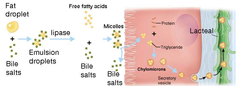 | |
Illustration of fat digestion. Click on image to enlarge
The bile acids remain in the intestinal lumen and travel to the terminal ileum where they are reabsorbed.
The micelles facilitate the transport of fatty acids and monoglycerides from the lumen across the epithelial membrane and into the cell. Once inside the enterocyte, fatty acids and monoglyceride are transported into the endoplasmic reticulum, where they are used to synthesise triglyceride. Beginning in the endoplasmic reticulum and continuing in the Golgi, triglyceride is packaged with cholesterol, lipoproteins and other lipids into particles called chylomicrons. Chylomicrons exit the cell by exocytosis across the basolateral membrane, they are too large to diffuse through capillaries and instead enter the lymphatics via endothelial gaps in the lacteals. The lymph is returned to the venous circulation via the lymphatic duct.
Vitamins
Fat soluble vitamins (A,D,E and K) enter the lymphatics as described above.Water soluble vitamins are typically absorbed via simple diffusion except vitamin B12 which is bound by intrinsic factor and absorbed in the terminal ileum via endocytosis.
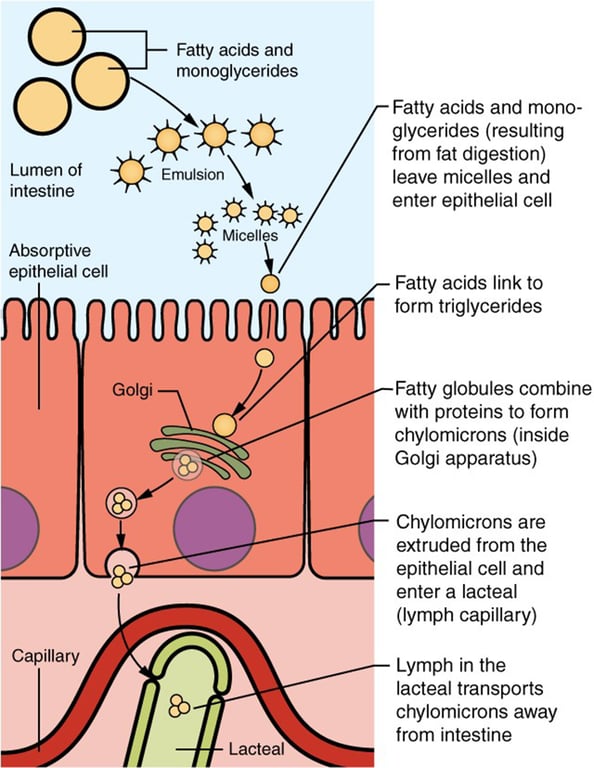 | |
Overview of fat handling by the small intestine. Click on image to enlarge