Cardiovascular physiology: Cardiac muscle
On this page
Cardiac muscle
The muscular tissue of the heart is called the myocardium and is responsible for generating forceful contraction of the atria and ventricles. The myocardium is composed of cardiac muscle cells called myocytes or cardiomyocytes.Structure
Cardiac muscles appear striated due to the orderly arrangement of thick (myosin) and thin (actin) filaments however they are not as orderly as skeletal muscle.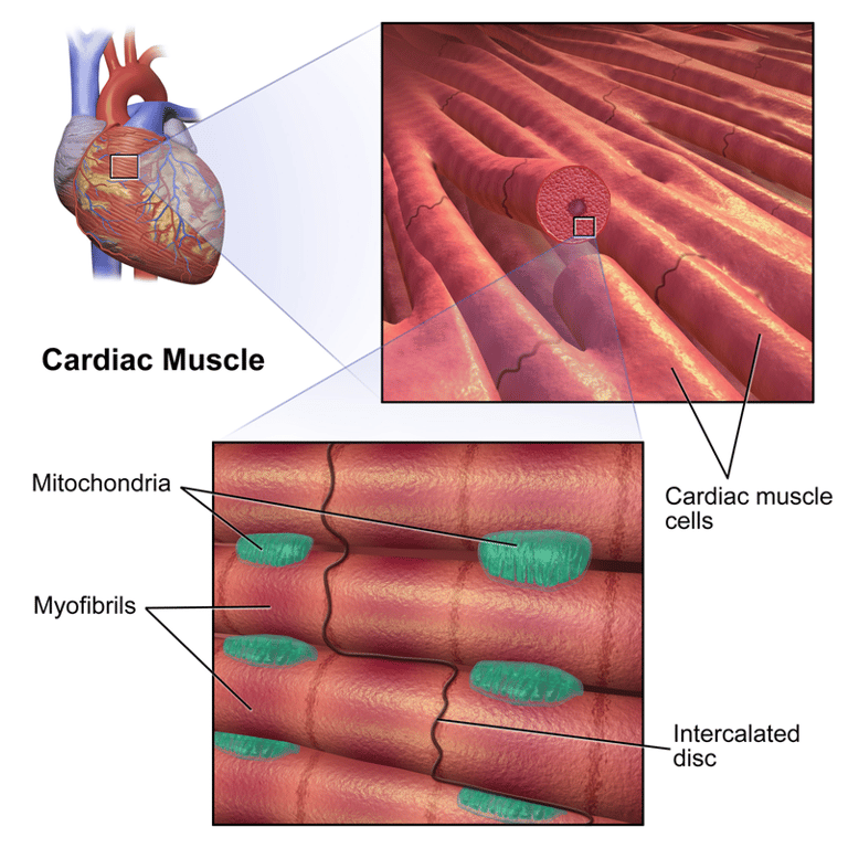 | |
Overview of appearance of cardiac muscle. Note multiple mitichondria. Connection to adjacent myocytes via intercalcated discs which contain gap junctions
Myocytes are branched and contain a single nucleus. Myocytes are rich in mitochondria to ensure a constant energy supply.
Electrical conduction & coupling
The heart generates its own rhythm to allow for co-ordinated pumping of the heart which requires synchronised contraction. Cardiac muscle is said to have inherent rhythmicity. Nerve impulses from the autonomic nervous system may modify the heart rate or force of contraction (chronotropic and inotropic effect) but they do not set the rhythm.Gap junctions between myocytes allow a low resistance pathway for depolarisation to spread rapidly between connected cells and for cardiac muscle to act as a functional syncytium (single unit).
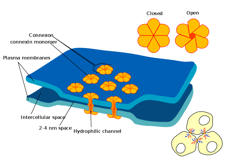 | |
Illustration of gap junctions communicating between myocytes connected via intercalated discs
Intercalated discs provide electrical connections between myocytes via gap junctions and structural attachment via
Gap junctions are composed of proteins called connexons
desmosomes
Gap junctions are composed of proteins called connexons
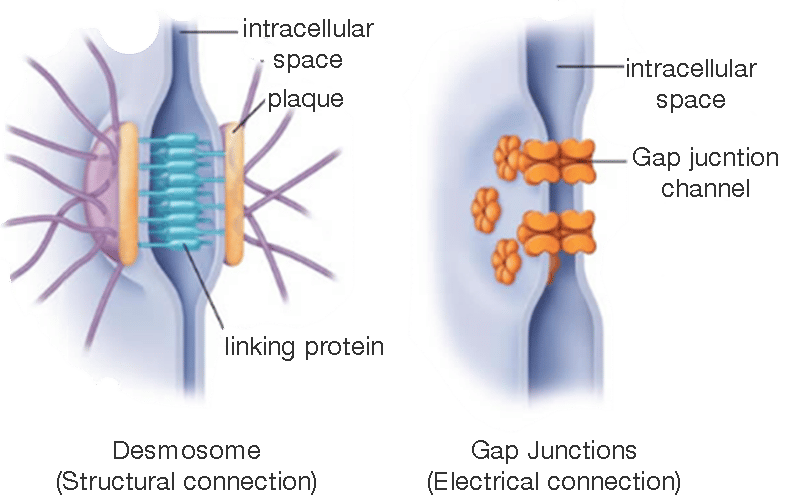 | |
Illustration of gap junctions and desmosomes communicating between myocytes connected via intercalated discs
Contraction of myocytes occurs due to a rise in intracellular Calcium ions. Excitation-contraction coupling
Cardiomyocytes contract when intracellular Ca2+ rises above 100nM. The rise in intracellular calcium occurs due to the following 2 mechanisms:- Release of calcium from sarcoplasmic reticulum (responsible for ~75% of the rise in Ca2+)
- Influx of calcium ions from outside of the cell (responsible for ~25% of the rise in Ca2+)
Action potentials then travel down extensions of the sarcolemma called T-tubules (transverse tubules).
1. Extracellular Ca2+ enters the myocyte via L-type calcium channels. This triggers the release of calcium from the sarcoplasmic reticulum (SR). This is termed calcium induced (or triggered) calcium release - CICR (or CTCR)
2. Calcium released from the SR binds to actin filaments facilitating contraction. Once contraction has completed calcium is actively and rapidly pumped back into the SR by the Ca2+-ATPase pump.
3. Ca2+ is also pumped out of the myocyte via Na+-Ca2+ exchangers in the cell membrane. This is relatively slow and occurs during diastole. If the heart rate increases, shortening diastole, the amount of calcium left in the cell is higher and the force of contraction is greater (This is known as the staircase or Treppe effect)
The video in the links shows an animated example of this.
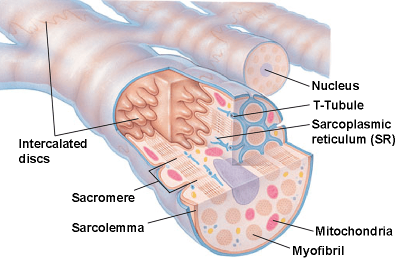 | |
Microscopic anatomy of cardiac muscle showing T-tubule network
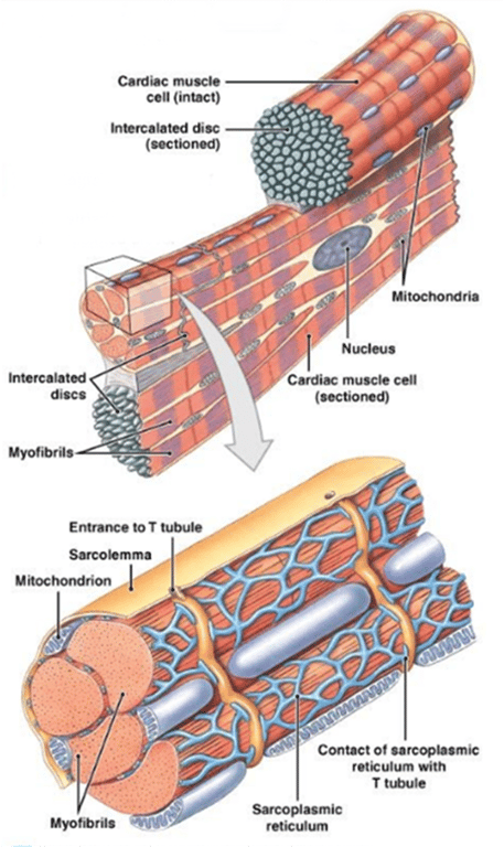 | |
Microscopic anatomy showing magnified section in context
Key differences between skeletal and cardiac muscles
| Cardiac muscle | Skeletal muscle | |
|---|---|---|
| Location | Heart only | Attached to bones via tendons |
| Stimulation | Involuntary | Voluntary |
| Nucleus | Single | Multinucleated |
| Appearance | Branching | Non-branched |
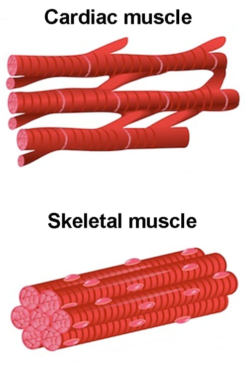 | |
Difference between cardiac and skeletal muscle. Note branching appearance of cardiac muscle