Respiratory physiology: Ventilation-perfusion mismatch
On this page
Ventilation and perfusion
Efficient gas exchange between air and blood at the alveolar interface are essential for oxygenation of the tissues and removal of carbon dioxide.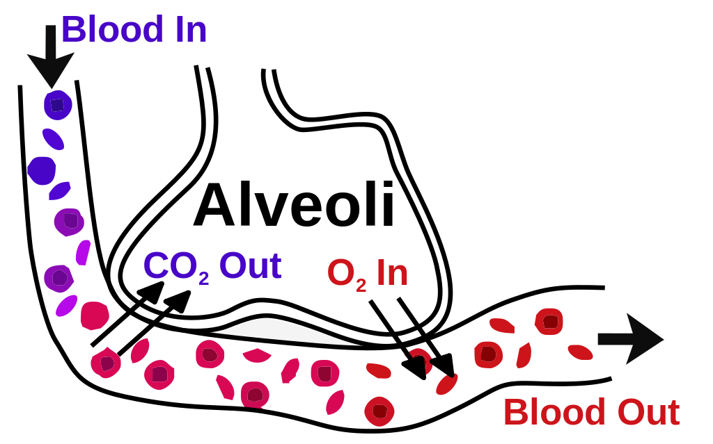 | |
Ventilation-perfusion ratio
The ventilation rate (V) refers to the volume of gas inhaled and exhaled from the lungs in one minute.
The ventilation rate (V) = tidal volume x respiratory rate
May be written as alveolar ventilation rate (Va)
May be written as alveolar ventilation rate (Va)
V typically equals 5-6 litres per minute.
Perfusion value (Q) of the lungs describes the volume of blood flowing through the pulmonary capillaries in one minute.
The ideal V/Q ratio is one.
Variations of ventilation and perfusion
The V/Q ratio varies throughout the lung. In healthy lungs the typical overall V/Q value is approximately 0.8V/Q ratio variation:
- Apex of lung: high V/Q (typically around 3)
- Base of lung: low V/Q (typically around 0.6)
Gravity is the principal cause of the V/Q gradient. In a standing individual both ventilation and perfusion increase going from the apex to the base however perfusion increases to a far greater degree than ventilation which lowers the V/Q ratio at the lung base. Patient position can therefore have a significant impact on V/Q
V/Q mismatch
V/Q mismatch maybe caused by inadequate ventilation or perfusion. The extremes of this are termed shunt and dead space.Extreme alterations of V/Q
- An area with perfusion but no ventilation (V/Q = zero) is termed shunt
- An area with ventilation but no perfusion (V/Q = approaching infinity) is termed dead space
In a true shunt where there is no ventilation increasing FiO2 will have no effect on blood oxygenation.
Both inadequate perfusion or ventilation can impede gas exchange and lead to hypoxaemia and hypercapnia.
Alveolar-arterial gradient (A-a gradient)
The A-a gradient measures the difference between the alveolar concentration (A) of oxygen and the arterial (a) concentration of oxygen. It is useful in diagnosing the source of hypoxaemia.
A-a gradient is calculated by:
- PAO2 (alveolar PO2) - PaO2 (arterial PO2)
Ideally there would be no A-a gradient as oxygen would reach equilibrium on both sides of the alveolar interface. In normal healthy individuals 'A' (PAO2) is higher than 'a' (PaO2) by at least 5-10 mmHg. This gradient exists due to physiological right-to-left shunting and the physiological V/Q variation (mismatch) in different lung zones described above primarily caused by gravity.
A V/Q mismatch will usually result in:
- Hypoxaemia
- Increased A-a gradient
Hypoxic vasoconstriction
If an area of the lung becomes inadequately ventilated (e.g. due to pneumonia) the resulting localised hypoxia stimulates vasoconstriction of arterioles supplying that lung area. This vasoconstriction diverts blood away from the under-ventilated area to better ventilated areas . This is known as shunting.