Lung root
The terms lung root (root of the lung) and hilum are sometimes used interchangeably. The lung hilum should be considered as the 2D area of the lung surface that receives the root of the lung whereas the root is the 3D pedicle that connects the lung to the mediastinum. The hilum marks the point where the parietal and visceral pleura meet and the pleural cavity starts and ends.
The root of the lung is a collection of structures that connect the lung to the mediastinum. The root of the lung is connected (by the structures that form it) to the heart and the trachea.
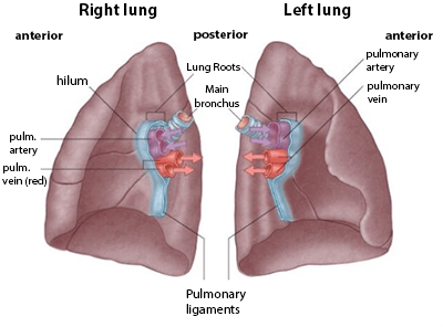
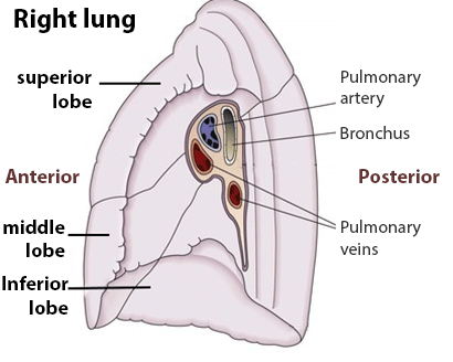
When considering the arrangement The root is often considered as having 4 main structures on each side (bronchus, pulmonary artery and 2 pulmonary veins - superior and inferior). The roots are arranged in a similar fashion from anterior to posterior on each side.
This means that the superior pulmonary vein are anterior, the pulmonary artery is in the middle, and the bronchus and bronchial vessels are behind. The second of the pulmonary veins is inferior to the bronchus. The anatomy is shown below:
Relations
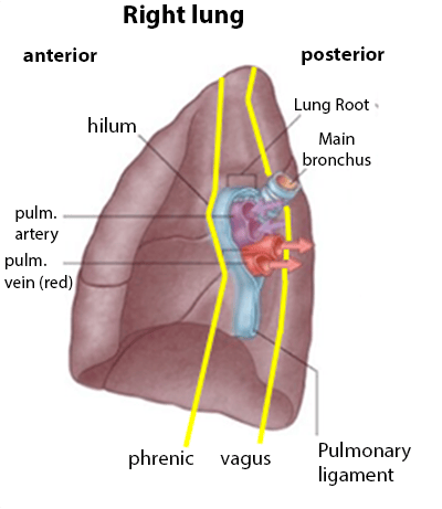
It is important to be aware of the anatomical relation of the lung root to the phrenic and vagus nerves.
Note the vagus gives off branches to supply the lungs along with sympathetic fibres from the sympathetic trunk. The phrenic nerve does not supply the lung (It gives off branches to supply the mediastinal pleura and pericardium on its way to supply the diaphragm)
The relations of the root to the phrenic and vagus nerves is shown below:
The root of the lung is a collection of structures that connect the lung to the mediastinum. The root of the lung is connected (by the structures that form it) to the heart and the trachea.
Structures forming the root of the lung
- Bronchus
- Pulmonary artery
- Pulmonary veins
- Bronchial arteries and veins
- Pulmonary plexuses of nerves
- Lymphatic vessels
When considering the arrangement The root is often considered as having 4 main structures on each side (bronchus, pulmonary artery and 2 pulmonary veins - superior and inferior). The roots are arranged in a similar fashion from anterior to posterior on each side.
This means that the superior pulmonary vein are anterior, the pulmonary artery is in the middle, and the bronchus and bronchial vessels are behind. The second of the pulmonary veins is inferior to the bronchus. The anatomy is shown below:
 | |
 | |
Relations
It is important to be aware of the anatomical relation of the lung root to the phrenic and vagus nerves.
Note the vagus gives off branches to supply the lungs along with sympathetic fibres from the sympathetic trunk. The phrenic nerve does not supply the lung (It gives off branches to supply the mediastinal pleura and pericardium on its way to supply the diaphragm)
The relations of the root to the phrenic and vagus nerves is shown below:
 | |