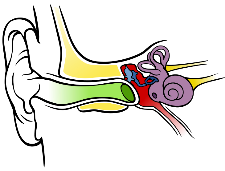
The ear can be divided anatomically into:
- External ear
- Middle ear
- Inner ear
Illustration of the different sections of the ear. External ear represented in green. Middle ear in red with ossicles in blue. Inner ear in purple
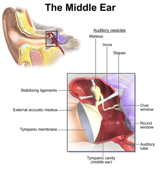
The middle ear is shown in more detail below:
Middle ear key points- Located in the petrous part of the temporal bone
- Air filled cavity between tympanic membrane and internal ear
- The cavity is sometimes split into the tympanic cavity and the epitympanic recess (the part of the cavity superior to the upper border of the tympanic membrane that houses the head of the malleus)
- Contains the ossicles (malleus, incus and stapes)
- The ossicles form communication from the external to internal/inner ear via the middle ear. The malleus is connected to the tympanic membrane and the stapes is connected to the oval window of the inner ear.
- The function of the middle ear is to convert air-borne vibrations to liquid-borne pulses
Important associated structures- Auditory (Eustachian) tube: connects the nasopharynx to the middle ear.
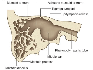
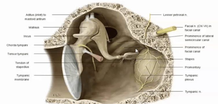
- Mastoid antrum: a collection of air-filled spaces in the mastoid process of the temporal bone posterior to the epitympanic recess that is connected to the middle ear via the aditus to mastoid antrum. The mucous membrane lining of the mastoid air cells is continuous with that of the tympanic membrane which is significant in that infections can easily spread between the two areas.
Images illustrating relationship between mastoid air cells and middle ear. Location of the aditus in the epitympanic recess of the middle ear