Thyroid: anatomy
On this page
The thyroid
The thyroid gland is positioned in the anterior neck spanning C5-T1 vertebral levels. It has two lobes connected by an isthmus.The isthmus
The isthmus lies over (anterior to) the 2nd-4th tracheal rings and posterior to the anterior jugular veins, sternothyroid and sternohyoid muscles.Arterial supply to the thyroid
The arterial supply to the thyroid is typically from the superior and inferior thyroid arteries. The superior thyroid artery arises from the external carotid artery. The inferior thyroid artery arises from the thyrocervical trunk.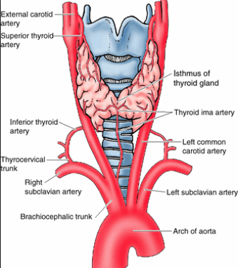 | |
Illustration of the arterial supply to the thyroid
Approximately 5-10% of the population will also have a patent thyroid ima artery. This is normally an embryonic remnant but in some it remains patent to provide arterial supply to the thyroid. It varies in origin with half a dozen possible source arteries but most commonly it arises from the brachiocephalic trunk
Venous drainage of the thyroid
Venous drainage is via the superior, middle and inferior thyroid veins. Superior and middle thyroid veins drain into the internal jugular vein. The inferior thyroid vein drains into the brachiocephalic vein.Lymph drainage of the thyroid
Lymph drainage is to the deep cervical lymph nodes.Anatomical relations of the thyroid
| Medial | Lateral | Posterior |
|---|---|---|
| Larynx Pharynx Trachea Oesophagus External laryngeal nerve Recurrent laryngeal nerve | Sternothyroid Sternohyoid Sternocleinomastoid Omohyoid | Parathyroids Carotid sheath Thoracic duct (left side) Inferior thyroid artery |
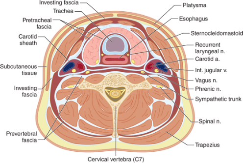 | |
Cross section of the neck showing anatomical relations
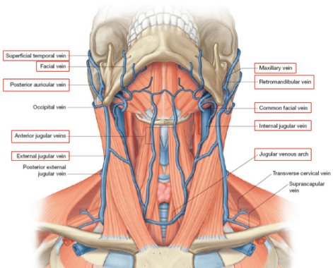 | |
Illustration of the superficial neck veins
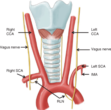 | |
Illustration of the recurrent laryngeal nerve anatomy