Orbit anatomy: foramina and fissures
Questions regarding orbital fractures and their sequelae have become a more common occurrence since the introduction of the 2015 curriculum. Candidates should be familiar with the structures passing through the superior and inferior orbital fissures and the optic canal. These are explained below.
Structures passing through optic canal
Structures passing through the superior orbital fissure
NERVES:
Structures passing through the inferior orbital fissure
Video highlighting the 3D anatomy of the orbital foramina
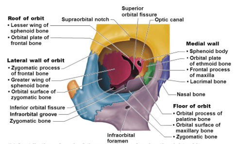 | |
Image showing the bone structure and foramina of the orbit
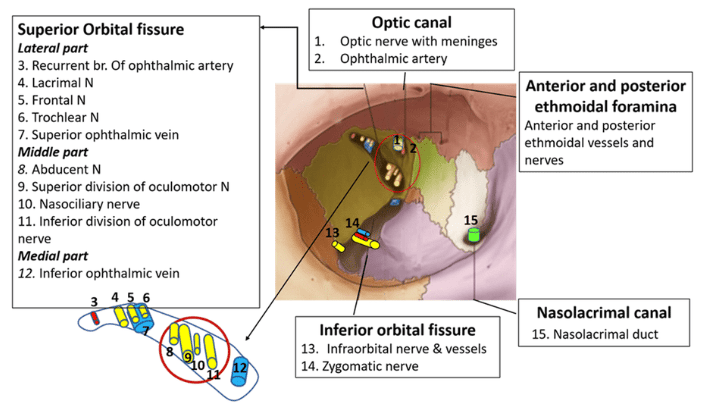 | |
Image summarising the structures passing through the fissures and foramina of the orbit
Structures passing through optic canal
- CN II, Optic nerve
- Ophthalmic artery
Structures passing through the superior orbital fissure
NERVES:
- CN III, oculomotor nerve (superior and inferior divisions)
- CN IV, trochlear nerve (IV)
- CN V1, trigeminal ophthalmic branch (lacrimal, frontal and nasociliary branches)
- CN VI, abducens nerve (VI)
- Sympathetic fibers from cavernous plexus
- Ophthalmic vein (superior and Inferior divisions) *inf. division also passes through inferior orbital fissure.
- Lacrimal artery (which is a branch of the ophthalmic artery. More specifically it is the recurrent meningeal branch)
Structures passing through the inferior orbital fissure
- Zygomatic nerve (branch of the maxillary nerve - CN V2)
- Infraorbital nerve (branch of the maxillary nerve - CN V2)
- Branches from the pterygopalatine ganglion.
- Ophthalmic vein (inferior branch)
- Infra-orbital artery and vein
Video highlighting the 3D anatomy of the orbital foramina