Parotid gland
Parotid gland overview
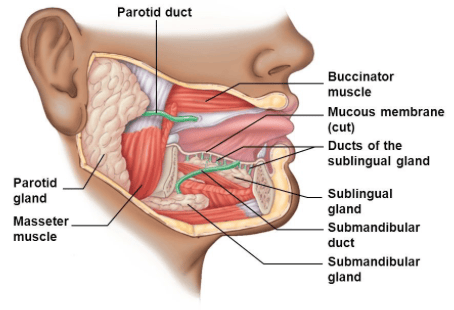
- Paired salivary glands
- Largest salivary glands
- Located in a hollow called the 'parotid region'
- It is covered by the parotid sheath which is a strong fibrous sheath which limits swelling and causes pain in parotid inflammation
- Drains via the parotid duct (Stensen duct)
 | |
Boundaries
| Boundary | Landmarks |
|---|---|
| Posteriorly | external ear and SCM muscle |
| Anteriorly | masseter muscle |
| Inferiorly | inferior border of the mandible |
| Superiorly | zygomatic arch |
Parotid duct
- Passes over the lateral surface of masseter then turns and pierces buccinator
- Passes forward from buccinator to open opposite the 2nd upper molar tooth
Innervation
- The parotid gland receives both sensory and autonomic innervation
- Sensory supply to the gland is from the auriculotemporal nerve and to the fascia/sheath via the great auricular nerve
- Secretomotor innervation (stimulation of saliva production) is via parasympathetic fibres of the glossopharyngeal nerve (CN IX). Parasympathetic innervation actually involves the CN IX fibres synapsing with the otic ganglion and these fibres are then carried by the auriculotemporal nerve to the parotid gland
- Saliva production is inhibited by stimulation of vasoconstriction via the sympathetic nervous system. These nerve fibres originate in the superior cervical ganglion and travel to the parotid via the external carotid artery nerve plexus.
Structures passing through the gland
- Facial nerve - divides into 5 terminal branches within the gland
- External carotid artery (ECA) - divides into 2 terminal branches (maxillary and superficial temporal arteries) within the gland
- Retromandibular vein - forms within the gland by the convergence of the superficial temporal and maxillary veins
- Parotid lymph nodes