Pleura
On this page
illustration showing pleura surface anatomy
The lungs are surrounded by two serous membranes called the visceral and pariteal pleurae (singular pleura). The visceral pleura is the deeper of the two and covers the lung parenchyma whilst the parietal pleura forms the outer layer. The space between the two is termed the pleural cavity and contains a small amount of fluid. The function of the fluid is to allow lubrication and provide surface tension.
For the exam detailed anatomy of the pleura is not a requirement of the college syllabus but candidates are expected to know the surface markings, how the pleura facilitate chest movement and the nerve supply to the pleura.
- Visceral pleura innervated by pulmonary plexus
- Visceral pleura lacks sensory innervation - parietal highly sensitive to pain
The lungs are surrounded by two serous membranes called the visceral and pariteal pleurae (singular pleura). The visceral pleura is the deeper of the two and covers the lung parenchyma whilst the parietal pleura forms the outer layer. The space between the two is termed the pleural cavity and contains a small amount of fluid. The function of the fluid is to allow lubrication and provide surface tension.
For the exam detailed anatomy of the pleura is not a requirement of the college syllabus but candidates are expected to know the surface markings, how the pleura facilitate chest movement and the nerve supply to the pleura.
Nerve supply
- Parietal pleura innervated by phrenic & intercostal nerves- Visceral pleura innervated by pulmonary plexus
- Visceral pleura lacks sensory innervation - parietal highly sensitive to pain
Surface markings
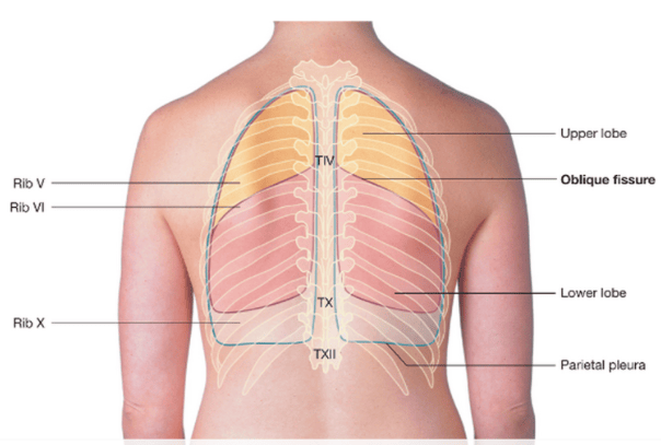
Candidates are also expected to know the function of the pleura in relation to facilitating chest movement and know the surface markings particularly in respect to safe chest drain insertion.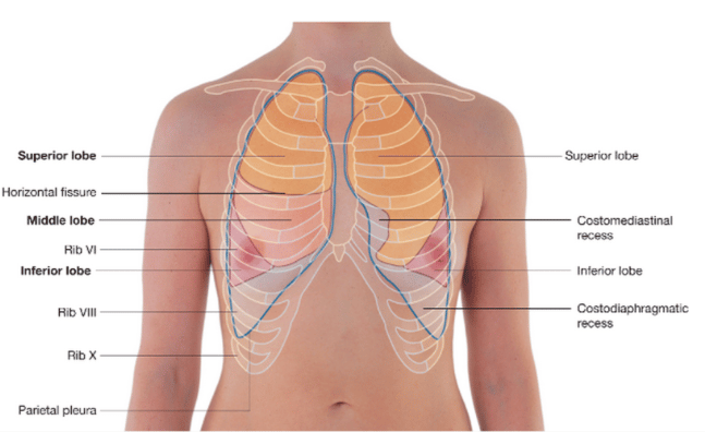 | |
| Wikipedia | |
 | |
| Wikipedia | |
Note superiorly the pleura form a curved apex behind the medial 1/3 of the clavicle with the dome extending above the clavicle. Note medially the left side of the pleura arches to accommodate the heart. Inferiorly you should know the boundaries of the pleura in the following: midclavicular line = 8th rib, midaxillary line= 10th rib, posteriorly runs in horizontal line intersecting the 12th rib and T12 vertebra