Skeletal muscle
On this page
There are 3 major muscle types:
Skeletal muscle is a striated muscle and is usually involved in movement of the body via the musculoskeletal system. It is typically under voluntary control via the somatic nervous system. Skeletal muscle uses approximately 25% of oxygen consumption at rest.
Muscle is composed of multiple parallel elongated multi-nucleated cells called myofibres or muscle fibres. Muscle fibres are 10-100 µm in diameter. Each myofibre is covered by a connective tissue called
Skeletal muscle is arranged in bundles of fibres called fasciculi (individual fasciculi are termed fasciculus or fascicle). Individual fasciculi are covered by perimysium,
Whole muscles are surrounded by a connective tissue called epimysium which extends beyond the muscle body to blend into the tendon.
Beneath the endomysium of each myofibre is sarcolemma. The sarcolemma is the excitable cell membrane of the muscle cell. The sarcolemma contains extensions that penetrate into the muscle cell called T-tubules.
Individual muscle fibres are composed of thousands of myofibrils. The myofibrils are composed of actin and myosin filaments arranged in repeating units called sarcomeres. The sarcomere is the basic functional units of the muscle fibre.
Myofibrils are only 1µm in diameter. They are organised in a parallel arrangement and separated from adjacent myofibrils by cytoplasm (the cytoplasm is called sarcoplasm). Each myofibril is composed of thick and thin filaments. The thick filaments are composed primarily of myosin and measure approximately 12nm in diameter. Thin filaments are composed of actin, tropomyosin and troponin and measure approximately 7nm in diameter.
The A-band describes the entire length of the thick myosin filament with its centre being the M-line (the M-line is a disc of cytoskeleton filaments that holds the myosin in position so each myosin is surrounded by 6 actin filaments in a hexagonal cross sectional structure). The paler central part of the A-band where there is only myosin filament and no overlapping actin is called the H-zone.
Sliding filaments
The sliding filament model or theory describes how muscles contract and relax. Thin filaments comprise strands of actin which contain globular binding sites for myosin. At rest these binding sites are covered by tropomyosin preventing myosin binding. The bulk of the thick filaments are comprised of myosin tails. Myosin heads project outward from the tails.
Large numbers of mitochondria and glycogen granules are located between myofibrils to provide the energy required for muscle contraction. T-Tubules are extensions of the plasma membrane (sarcolemma) that invaginate into muscle cells. The smooth endoplasmic reticulum of muscle cells is called sarcoplasmic reticulum and enlarges near T-tubules to form terminal cisternae. The sarco-endoplasmic reticulum Ca2+-ATPase (SERCA) pump transports Ca2+ into the sarcoplasmic reticulum (SR). The primary function of the sarcoplasmic reticulum (SR) is to store calcium and release it for muscle contraction. Located within the SR is a protein called calsequestrin. This protein can bind to around 50 Ca2+ ions and allows the SR to store vast quantities of calcium.
Contraction of skeletal muscle is brought about by the release of Ca2+ from the SR. Ca2+ binds to troponin on the thin filaments and this causes a conformational change that shifts tropomyosin and exposes actin binding sites (shown in the video below). This allows myosin to form a cross bridge with actin.
Myosin prepares for binding to actin by tilting its head. It does this by utilising ATP (which undergoes hydrolysis via the enzyme myosin ATPase, and is converted to a phosphate group and ADP). The myosin head is said to be energised and is bound to ADP.
After binding to actin (forming a crossbridge) the myosin head then changes shape again when it tilts (or rotates) forward pulling on the actin filament. This is referred to as the power stroke. Myosin pulling on actin causes the muscle fibre to shorten. This is shown in the video below.
Following the power stroke ADP is released from the myosin head and this allows the myosin head to dissociate form the actin filament and bind with another ATP molecule.
- Skeletal muscle
- Cardiac muscle
- Smooth muscle
Skeletal muscle is a striated muscle and is usually involved in movement of the body via the musculoskeletal system. It is typically under voluntary control via the somatic nervous system. Skeletal muscle uses approximately 25% of oxygen consumption at rest.
Skeletal muscle composition overview
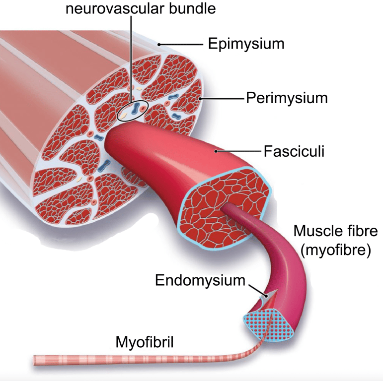 | |
Overview of muscle structure. click on image to enlarge
Muscle is composed of multiple parallel elongated multi-nucleated cells called myofibres or muscle fibres. Muscle fibres are 10-100 µm in diameter. Each myofibre is covered by a connective tissue called
endomysium
Skeletal muscle is arranged in bundles of fibres called fasciculi (individual fasciculi are termed fasciculus or fascicle). Individual fasciculi are covered by perimysium,
Whole muscles are surrounded by a connective tissue called epimysium which extends beyond the muscle body to blend into the tendon.
Skeletal muscle fibre structure
Beneath the endomysium of each myofibre is sarcolemma. The sarcolemma is the excitable cell membrane of the muscle cell. The sarcolemma contains extensions that penetrate into the muscle cell called T-tubules.
T-tubules permit rapid transmission of the action potential into the cell, and also play an important role in regulating cellular calcium concentration.
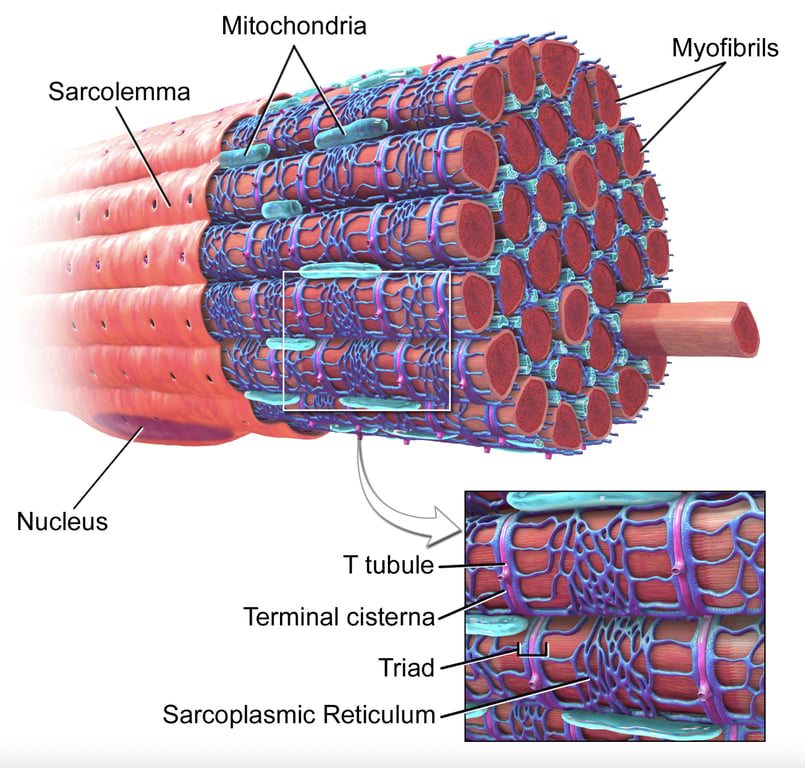 | |
Diagram illustrating skeletal muscle highlighting sarcolemma. click on image to enlarge
Muscle fibres are cylindrical and are rich in mitochondria to fulfil the high energy demands of the myocyte.
Individual muscle fibres are composed of thousands of myofibrils. The myofibrils are composed of actin and myosin filaments arranged in repeating units called sarcomeres. The sarcomere is the basic functional units of the muscle fibre.
Myofibrils are only 1µm in diameter. They are organised in a parallel arrangement and separated from adjacent myofibrils by cytoplasm (the cytoplasm is called sarcoplasm). Each myofibril is composed of thick and thin filaments. The thick filaments are composed primarily of myosin and measure approximately 12nm in diameter. Thin filaments are composed of actin, tropomyosin and troponin and measure approximately 7nm in diameter.
Sarcomeres
Myofibrils are divided longitudinally into sarcomeres. Sarcomeres are separated from each other by Z-discs or Z-lines. The Z-lines are the anchoring points for thin actin filaments. The I-band extends from the Z-line and is the area containing only actin thin filaments i.e. it is NOT superimposed by thick filaments (myosin).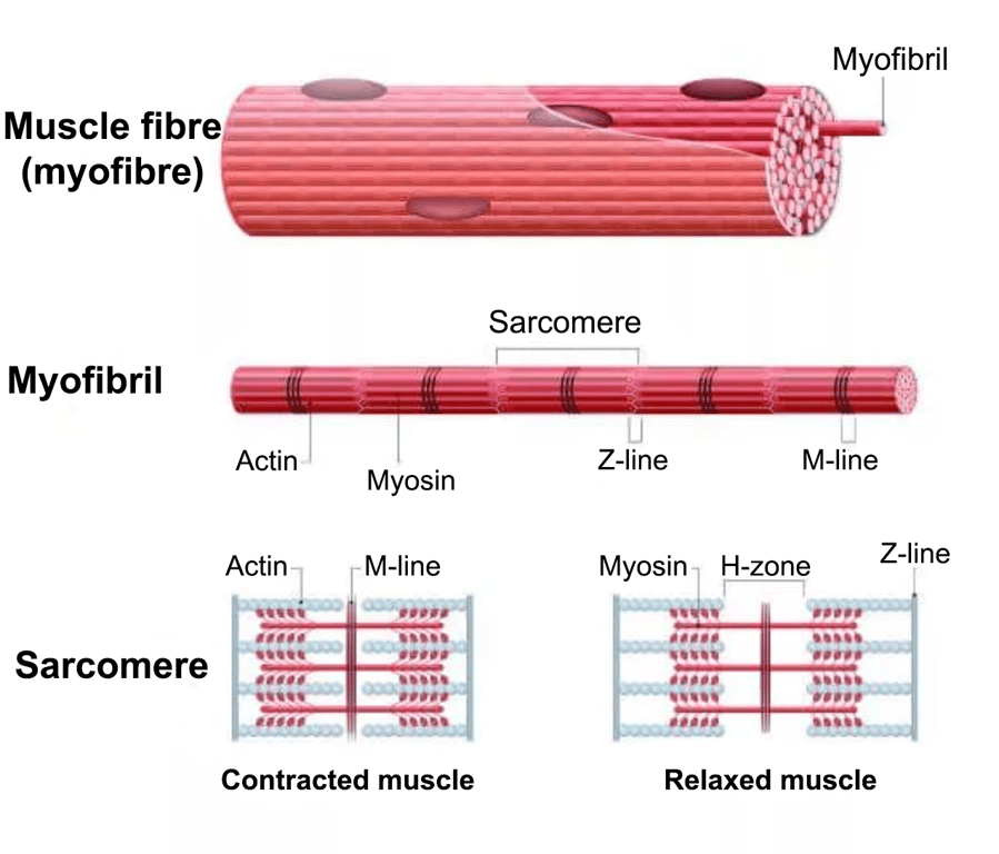 | |
Image showing relationship between myofibres, myofibril and sarcomere. click on image to enlarge
The A-band describes the entire length of the thick myosin filament with its centre being the M-line (the M-line is a disc of cytoskeleton filaments that holds the myosin in position so each myosin is surrounded by 6 actin filaments in a hexagonal cross sectional structure). The paler central part of the A-band where there is only myosin filament and no overlapping actin is called the H-zone.
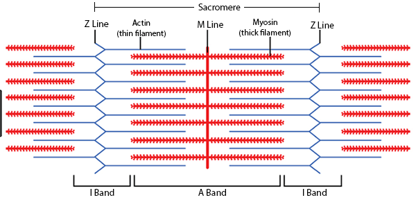 | |
Skeletal muscle bands illustrated. click on image to enlarge
Sliding filaments
The sliding filament model or theory describes how muscles contract and relax. Thin filaments comprise strands of actin which contain globular binding sites for myosin. At rest these binding sites are covered by tropomyosin preventing myosin binding. The bulk of the thick filaments are comprised of myosin tails. Myosin heads project outward from the tails.
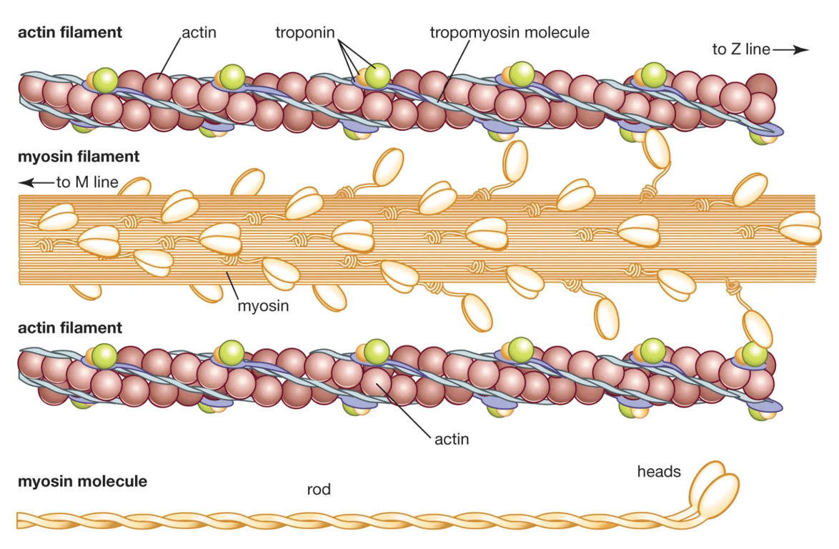 | |
Actin and myosin filaments illustrated. click on image to enlarge
Large numbers of mitochondria and glycogen granules are located between myofibrils to provide the energy required for muscle contraction. T-Tubules are extensions of the plasma membrane (sarcolemma) that invaginate into muscle cells. The smooth endoplasmic reticulum of muscle cells is called sarcoplasmic reticulum and enlarges near T-tubules to form terminal cisternae. The sarco-endoplasmic reticulum Ca2+-ATPase (SERCA) pump transports Ca2+ into the sarcoplasmic reticulum (SR). The primary function of the sarcoplasmic reticulum (SR) is to store calcium and release it for muscle contraction. Located within the SR is a protein called calsequestrin. This protein can bind to around 50 Ca2+ ions and allows the SR to store vast quantities of calcium.
Contraction is maintained as long as Ca2+ levels remain high. The contraction time is therefore dependent on the rate at which SERCA pumps Ca2+ back into the SR
Contraction of skeletal muscle is brought about by the release of Ca2+ from the SR. Ca2+ binds to troponin on the thin filaments and this causes a conformational change that shifts tropomyosin and exposes actin binding sites (shown in the video below). This allows myosin to form a cross bridge with actin.
Myosin prepares for binding to actin by tilting its head. It does this by utilising ATP (which undergoes hydrolysis via the enzyme myosin ATPase, and is converted to a phosphate group and ADP). The myosin head is said to be energised and is bound to ADP.
After binding to actin (forming a crossbridge) the myosin head then changes shape again when it tilts (or rotates) forward pulling on the actin filament. This is referred to as the power stroke. Myosin pulling on actin causes the muscle fibre to shorten. This is shown in the video below.
Following the power stroke ADP is released from the myosin head and this allows the myosin head to dissociate form the actin filament and bind with another ATP molecule.
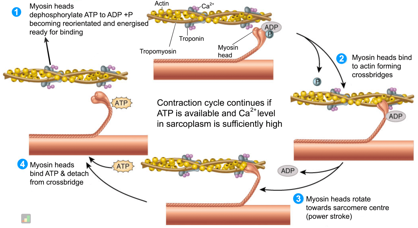 | |
Summary graphic of the sliding filament model. click on image to enlarge