Ulcerative colitis
Ulcerative colitis (UC) is a form of inflammatory bowel disease. Inflammation always starts at rectum (hence it is the most common site for UC), never spreads beyond ileocaecal valve and is continuous. The peak incidence of ulcerative colitis is in people aged 15-25 years and in those aged 55-65 years.
The initial presentation is usually following insidious and intermittent symptoms. Features include:
Questions regarding the 'extra-intestinal' features of inflammatory bowel disease are common:
Pathology
Barium enema
The initial presentation is usually following insidious and intermittent symptoms. Features include:
- bloody diarrhoea
- urgency
- tenesmus
- abdominal pain, particularly in the left lower quadrant
- extra-intestinal features (see below)
Questions regarding the 'extra-intestinal' features of inflammatory bowel disease are common:
| Common to both Crohn's disease (CD) and Ulcerative colitis (UC) | Notes | |
|---|---|---|
| Related to disease activity | Arthritis: pauciarticular, asymmetric Erythema nodosum Episcleritis Osteoporosis | Arthritis is the most common extra-intestinal feature in both CD and UC Episcleritis is more common in CD |
| Unrelated to disease activity | Arthritis: polyarticular, symmetric Uveitis Pyoderma gangrenosum Clubbing Primary sclerosing cholangitis | Primary sclerosing cholangitis is much more common in UC Uveitis is more common in UC |
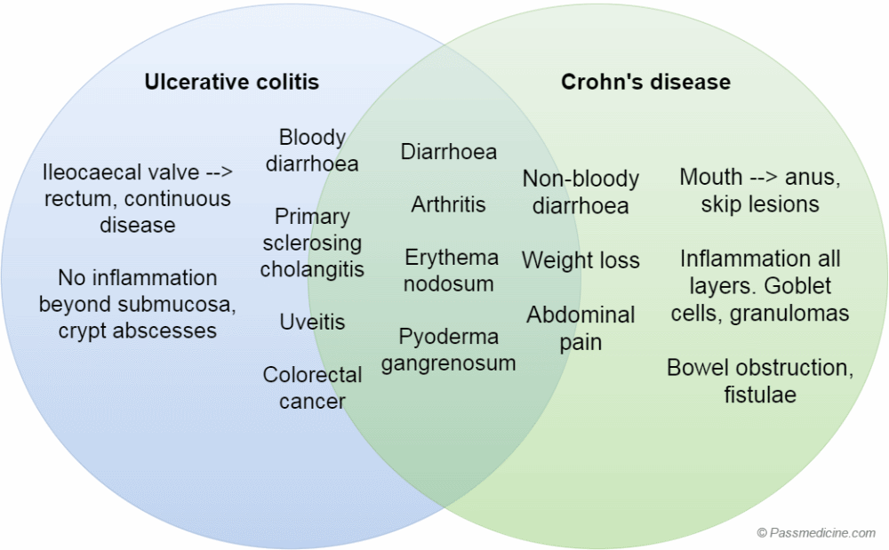 | |
Venn diagram showing shared features and differences between ulcerative colitis and Crohn's disease. Note that whilst some features are present in both, some are much more common in one of the conditions, for example colorectal cancer in ulcerative colitis
Pathology
- red, raw mucosa, bleeds easily
- no inflammation beyond submucosa (unless fulminant disease)
- widespread ulceration with preservation of adjacent mucosa which has the appearance of polyps ('pseudopolyps')
- inflammatory cell infiltrate in lamina propria
- neutrophils migrate through the walls of glands to form crypt abscesses
- depletion of goblet cells and mucin from gland epithelium
- granulomas are infrequent
Barium enema
- loss of haustrations
- superficial ulceration, 'pseudopolyps'
- long standing disease: colon is narrow and short -'drainpipe colon'
 | |
| © Image used on license from Radiopaedia | |
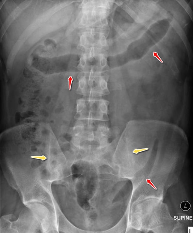
Abdominal x-ray from a patient with ulcerative colitis showing lead pipe appearance of the colon (red arrows). Ankylosis of the left sacroiliac joint and partial ankylosis on the right (yellow arrow), reinforcing the link with sacroilitis.
 | |
| © Image used on license from Radiopaedia | |
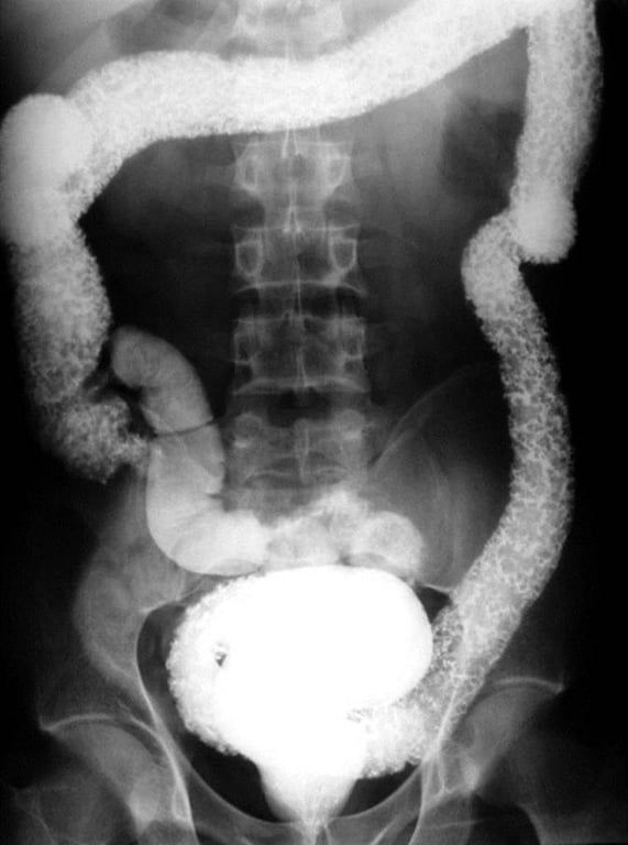
Barium enema from a patient with ulcerative colitis. The whole colon, without skips is affected by an irregular mucosa with loss of normal haustral markings.