Cardiovascular physiology: Cardiac action potential
On this page
Cardiac action potential
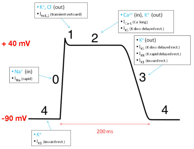 | |
Ventricular myocyte depolarisation. Click on image to enlarge
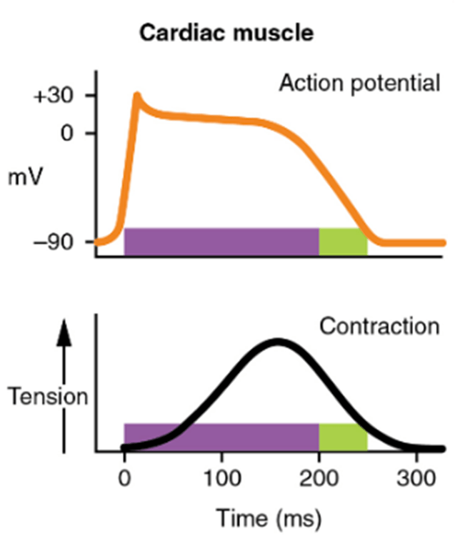 | |
Note the relationship of depolarisation to muscle contraction. Na+ influx causes the initial depolarisation but it is the influx of Ca2+ that leads to muscle contraction towards the end of phase 2
| Phase | Description | Mechanism |
|---|---|---|
| 0 | Rapid depolarisation | Rapid sodium influx These channels automatically deactivate after a few ms |
| 1 | Early repolarisation | Efflux of potassium |
| 2 | Plateau | Slow influx of calcium |
| 3 | Final repolarisation | Efflux of potassium |
| 4 | Restoration of ionic concentrations | Resting potential is restored by Na+/K+ ATPase There is slow entry of Na+ into the cell decreasing the potential difference until the threshold potential is reached, triggering a new action potential |
NB cardiac muscle remains contracted 10-15 times longer than skeletal muscle
Conduction velocity
| Site | Speed |
|---|---|
| Atrial conduction | Spreads along ordinary atrial myocardial fibres at 1 m/sec |
| AV node conduction | 0.05 m/sec |
| Ventricular conduction | Purkinje fibres are of large diameter and achieve velocities of 2-4 m/sec (this allows a rapid and coordinated contraction of the ventricles |
The SA node
Note that action potentials are not the same in all myocytes. Atrial myocytes have similar graphs to ventricular myocytes (as shown above) but the atrial action potential is shorter than the ventricular action potential. The plateau phase in the atrial action potential is also at a lower potential than that seen in the ventricle.
Pacemaker cells have more significant differences. The SA node lacks fast Na channels. Depolarisation of the SA node myocytes is via influx of Ca2+ ions via slow Ca2+ channels (rather than influx of sodium via fast Na channels). This results in slower depolarisation.
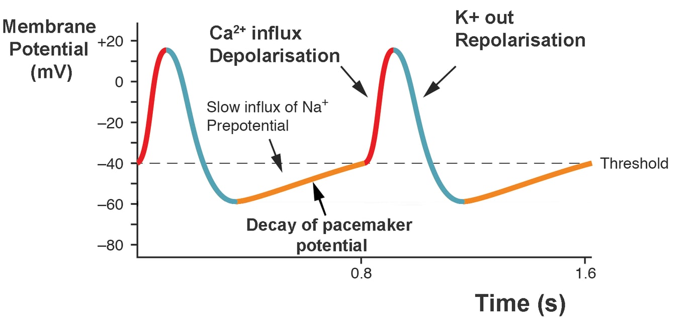 | |
SA pacemaker depolarisation. Click on image to enlarge
Summary of resting and threshold potentials for pacemaker and non-pacemaker cells
| Site | Resting potential | Threshold potential |
|---|---|---|
| SAN | -60mV | -40mV |
| Ventricular and atrial myocytes | -90mV | -70mV |