ECG: left bundle branch block
On this page
The diagram below shows the typical features of left bundle branch block (LBBB):
One of the most common ways to remember the difference between LBBB and RBBB is WiLLiaM MaRRoW
Causes of LBBB
New LBBB is always pathological and may be a sign of a myocardial infarction. Diagnosing a myocardial infarction for patients with pre-existing LBBB is difficult. The Sgarbossa criteria can help with this. See below:
Interpretation: A score of <3 is not considered diagnostic of acute MI but also does not rule out MI. Scores ≥ 3 have a specificity of 90% for diagnosing acute MI.
The Sgarbossa criteria were modified by Dr Smith (of Dr Smiths ECG blog fame) to become the modified Sgarbossa's criteria. In these criteria the third criteria in the table above has the original absolute 5 mm threshold replaced with a proportion: ST elevation/S-wave amplitude of ≤ -0.25.
In the modified criteria if any of the criterion are positive this supports a diagnosis of acute MI.
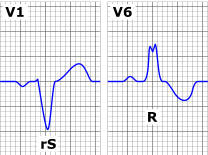 | |
One of the most common ways to remember the difference between LBBB and RBBB is WiLLiaM MaRRoW
- in LBBB there is a 'W' in V1 and a 'M' in V6
- in RBBB there is a 'M' in V1 and a 'W' in V6
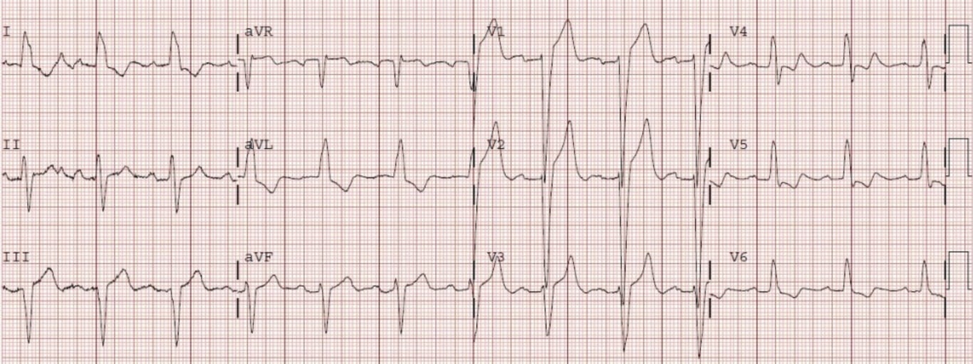 | |
ECG showing typical features of LBBB. click on image to enlarge
Causes of LBBB
- ischaemic heart disease
- hypertension
- aortic stenosis
- cardiomyopathy
- rare: idiopathic fibrosis, digoxin toxicity, hyperkalaemia
New LBBB is always pathological and may be a sign of a myocardial infarction. Diagnosing a myocardial infarction for patients with pre-existing LBBB is difficult. The Sgarbossa criteria can help with this. See below:
Sgarbossa criteria
| Criteria | Points if criteria met |
|---|---|
| Concordant ST elevation > 1mm in leads with a positive QRS complex | 5 |
| Concordant ST depression > 1 mm in V1-V3 | 3 |
| Excessively discordant ST elevation ≥5 mm in leads with a negative QRS) | 2 |
Interpretation: A score of <3 is not considered diagnostic of acute MI but also does not rule out MI. Scores ≥ 3 have a specificity of 90% for diagnosing acute MI.
The Sgarbossa criteria were modified by Dr Smith (of Dr Smiths ECG blog fame) to become the modified Sgarbossa's criteria. In these criteria the third criteria in the table above has the original absolute 5 mm threshold replaced with a proportion: ST elevation/S-wave amplitude of ≤ -0.25.
In the modified criteria if any of the criterion are positive this supports a diagnosis of acute MI.
Modified Sgarbossa criteria
| Criteria |
|---|
| Concordant ST elevation > 1mm in leads with a positive QRS complex |
| Concordant ST depression > 1 mm in V1-V3 |
| ST Elevation at the J-point, relative to QRS onset, is at least 1 mm AND has an amplitude at least 25% of the preceding S-wave |