Mitral stenosis
It is said that the causes of mitral stenosis are rheumatic fever, rheumatic fever and rheumatic fever. Rarer causes that may be seen in the exam include mucopolysaccharidoses, carcinoid and endocardial fibroelastosis
Features
Features of severe MS
Chest x-ray
Echocardiography
Features
- mid-late diastolic murmur (best heard in expiration)
- loud S1, opening snap
- low volume pulse
- malar flush
- atrial fibrillation
Features of severe MS
- length of murmur increases
- opening snap becomes closer to S2
Chest x-ray
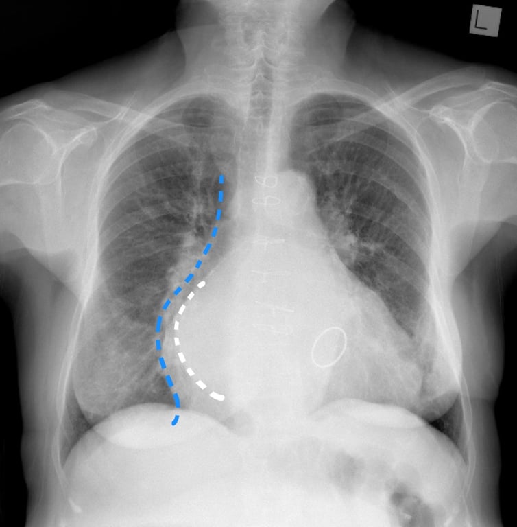
- left atrial enlargement may be seen
Echocardiography
- the normal cross sectional area of the mitral valve is 4-6 sq cm. A 'tight' mitral stenosis implies a cross sectional area of < 1 sq cm
 | |
| © Image used on license from Radiopaedia | |
Chest x-ray from a patient with mitral stenosis. This patient has had a sternotomy and a prosthetic mitral valve. There is splaying of the carina with elevation of the left main bronchus, a double right heart border and cardiomegaly. The features are those of left atrial enlargement. Although the entire heart is enlarged, a double contour is seen through the right side of the heart. The more medial line is the enlarged left atrium (white dotted line) and the heart heart border is more lateral (blue dotted line).