Stroke: a very basic introduction
Strokes represent an important cause of morbidity and mortality. In the UK alone there are over 150,000 strokes per year, with over 1.2 million stroke survivors. Stroke is the fourth largest cause of death in the UK and kills twice as many women than breast cancer each year.
The prevention and treatment of strokes has undergone significant changes over the past decade. What was previously considered a devastating but untreatable condition is now viewed more as a 'brain attack', a condition which requires emergency assessment to see if patients may benefit from new treatments such as thrombolysis.
A stroke (also known as cerebrovascular accident,CVA) represents a sudden interruption in the vascular supply of the brain. Remember that neural tissue is completely dependent on aerobic metabolism so any problem with oxygen supply can quickly lead to irreversible damage.
There are two main types of strokes:
The table below shows the basic differences:
Symptoms and signs
Stroke is defined by the World Health Organization as a clinical syndrome consisting of 'rapidly developing clinical signs of focal (at times global) disturbance of cerebral function, lasting more than 24 hours or leading to death with no apparent cause other than that of vascular origin'. In contrast, with a TIA the symptoms and signs resolve within 24 hours.
Features include:
Cerebral hemisphere infarcts may have the following symptoms:
Brainstem infarction
Lacunar infarcts
One formal classification system that is sometimes used is the Oxford Stroke Classification (also known as the Bamford Classification), which classifies strokes based on the initial symptoms. A summary is as follows:
The following criteria should be assessed:
Whilst symptoms alone cannot be used to differentiate haemorrhagic from ischaemic strokes, patients who've suffered haemorrhages are more likely to have:
Over recent years there has been a public health campaign to raise awareness of stroke symptoms. The FAST campaign uses the following mnemonic:
Investigations
Patients with suspected stroke need to have emergency neuroimaging. The main cause for urgency is to see whether a patient may be suitable for thrombolytic therapy to treat early ischaemic strokes. The two types of neuroimaging used in this setting are:
Management
Ischaemic strokes
Urgent neuroimaging classifies the stroke as either ischaemic or haemorrhagic. If the stroke is ischaemic, and certain criteria are met, the patient should be offered thrombolysis. Example criteria include:
Once haemorrhagic stroke has been excluded patients should be given aspirin 300mg as soon as possible and antiplatelet therapy should be continued.
Remember with TIAs the, by definition, symptoms last less than 24 hours although in the vast majority of cases the duration is much shorter, typically 1 hour or so. For this reason most patients symptoms will have resolved before they see a doctor.
The treatment of TIAs focuses around reducing the risk of further ischaemic events. NICE currently advocate using a risk based approach based around the ABCD2 prognostic score:
This gives a total score ranging from 0 to 7. People who have had a suspected TIA who are at a higher risk of stroke (that is, with an ABCD2 score of 4 or above) should have:
If the ABCD2 risk score is 3 or below:
People with crescendo TIAs (two or more episodes in a week) should be treated as being at high risk of stroke, even though they may have an ABCD2 score of 3 or below.
If imaging confirms a haemorrhagic stroke neurosurgical consultation should be considered for advice on further management. The vast majority of patients however are not suitable for surgical intervention. Management is therefore supportive as per haemorrhagic stroke. Anticoagulants (e.g. warfarin) and antithrombotic medications (e.g. clopidogrel) should be stopped to minimise further bleeding. If a patient is anticoagulated this should be reversed as quickly as possible. Trials have shown improved outcomes in patients who have their blood pressure lowered acutely and this is now part of many protocols for haemorrhagic strokes.
The prevention and treatment of strokes has undergone significant changes over the past decade. What was previously considered a devastating but untreatable condition is now viewed more as a 'brain attack', a condition which requires emergency assessment to see if patients may benefit from new treatments such as thrombolysis.
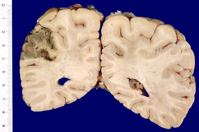 | |
| © Image used on license from PathoPic | |
Pathological specimen showing the results of an ischaemic stroke to the occipito-parietal region of the cerebrum. Note there has been some secondary haemorrhage in the affected area.
What is a stroke?
A stroke (also known as cerebrovascular accident,CVA) represents a sudden interruption in the vascular supply of the brain. Remember that neural tissue is completely dependent on aerobic metabolism so any problem with oxygen supply can quickly lead to irreversible damage.
There are two main types of strokes:
- ischaemic: these can be further subdivided between into episodes which last greater than 24 hours (termed an ischaemic stroke) and episodes where symptoms and signs last less than 24 hours (transient ischaemic attacks, TIAs, sometimes termed 'mini-strokes' by patients)
- haemorrhagic
The table below shows the basic differences:
| Ischaemic | Haemorrhagic | |
|---|---|---|
| Essential problem | 'Blockage' in the blood vessel stops blood flow | Blood vessel 'bursts' leading to reduction in blood flow |
| Proportion of strokes | 85% | 15% |
| Subtypes | Thrombotic stroke
Embolic stroke
| Intracerebral haemorrhage
Subarachnoid haemorrhage
|
| Risk factors | General risk factors for cardiovascular disease
Risk factors for cardioembolism
| Risk factors
|
Symptoms and signs
Stroke is defined by the World Health Organization as a clinical syndrome consisting of 'rapidly developing clinical signs of focal (at times global) disturbance of cerebral function, lasting more than 24 hours or leading to death with no apparent cause other than that of vascular origin'. In contrast, with a TIA the symptoms and signs resolve within 24 hours.
Features include:
- motor weakness
- speech problems (dysphasia)
- swallowing problems
- visual field defects (homonymous hemianopia)
- balance problems
Cerebral hemisphere infarcts may have the following symptoms:
- contralateral hemiplegia: initially flaccid then spastic
- contralateral sensory loss
- homonymous hemianopia
- dysphasia
Brainstem infarction
- may result in more severe symptoms including quadriplegia and lock-in-syndrome
Lacunar infarcts
- small infarcts around the basal ganglia, internal capsule, thalamus and pons
- this may result in pure motor, pure sensory, mixed motor and sensory signs or ataxia
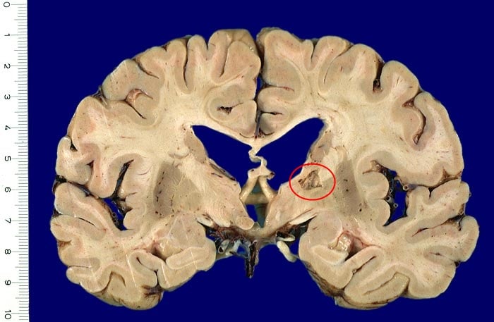 | |
| © Image used on license from PathoPic | |
An example of a lacunar infarct affecting the internal capsule.
One formal classification system that is sometimes used is the Oxford Stroke Classification (also known as the Bamford Classification), which classifies strokes based on the initial symptoms. A summary is as follows:
The following criteria should be assessed:
- 1. unilateral hemiparesis and/or hemisensory loss of the face, arm & leg
- 2. homonymous hemianopia
- 3. higher cognitive dysfunction e.g. dysphasia
| Stroke type | Notes |
|---|---|
| Total anterior circulation infarcts (TACI, c. 15%) |
|
| Partial anterior circulation infarcts (PACI, c. 25%) |
|
| Lacunar infarcts (LACI, c. 25%) |
|
| Posterior circulation infarcts (POCI, c. 25%) |
|
Whilst symptoms alone cannot be used to differentiate haemorrhagic from ischaemic strokes, patients who've suffered haemorrhages are more likely to have:
- decrease in the level of consciousness: seen in up to 50% of patients with a haemorrhagic stroke
- headache is also much more common in haemorrhagic stroke
- nausea and vomiting is also common
- seizures occur in up to 25% of patients
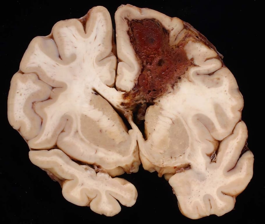 | |
| © Image used on license from PathoPic | |
Pathological specimen showing the consequence of an intracerebral haemorrhage
Over recent years there has been a public health campaign to raise awareness of stroke symptoms. The FAST campaign uses the following mnemonic:
- Face - 'Has their face fallen on one side? Can they smile?'
- Arms - 'Can they raise both arms and keep them there?'
- Speech - 'Is their speech slurred?'
- Ttime - 'Time to call 999 if you see any single one of these signs.'
Investigations
Patients with suspected stroke need to have emergency neuroimaging. The main cause for urgency is to see whether a patient may be suitable for thrombolytic therapy to treat early ischaemic strokes. The two types of neuroimaging used in this setting are:
- CT
- MRI
Management
Ischaemic strokes
Urgent neuroimaging classifies the stroke as either ischaemic or haemorrhagic. If the stroke is ischaemic, and certain criteria are met, the patient should be offered thrombolysis. Example criteria include:
- patients present with 4.5 hours of onset of stroke symptoms
- the patient has not had a previous intracranial haemorrhage, uncontrolled hypertension, pregnant etc
Once haemorrhagic stroke has been excluded patients should be given aspirin 300mg as soon as possible and antiplatelet therapy should be continued.
Transient ischaemic attacks
Remember with TIAs the, by definition, symptoms last less than 24 hours although in the vast majority of cases the duration is much shorter, typically 1 hour or so. For this reason most patients symptoms will have resolved before they see a doctor.
The treatment of TIAs focuses around reducing the risk of further ischaemic events. NICE currently advocate using a risk based approach based around the ABCD2 prognostic score:
| Criteria | Points | |
|---|---|---|
| A | Age >= 60 years | 1 |
| B | Blood pressure >= 140/90 mmHg | 1 |
| C | Clinical features - Unilateral weakness - Speech disturbance, no weakness | 2 1 |
| D | Duration of symptoms - > 60 minutes - 10-59 minutes | 2 1 |
| Patient has diabetes | 1 |
This gives a total score ranging from 0 to 7. People who have had a suspected TIA who are at a higher risk of stroke (that is, with an ABCD2 score of 4 or above) should have:
- aspirin (300 mg daily) started immediately
- specialist assessment and investigation within 24 hours of onset of symptoms
- measures for secondary prevention introduced as soon as the diagnosis is confirmed, including discussion of individual risk factors
If the ABCD2 risk score is 3 or below:
- specialist assessment within 1 week of symptom onset, including decision on brain imaging
- if vascular territory or pathology is uncertain, refer for brain imaging
People with crescendo TIAs (two or more episodes in a week) should be treated as being at high risk of stroke, even though they may have an ABCD2 score of 3 or below.
Haemorrhagic strokes
If imaging confirms a haemorrhagic stroke neurosurgical consultation should be considered for advice on further management. The vast majority of patients however are not suitable for surgical intervention. Management is therefore supportive as per haemorrhagic stroke. Anticoagulants (e.g. warfarin) and antithrombotic medications (e.g. clopidogrel) should be stopped to minimise further bleeding. If a patient is anticoagulated this should be reversed as quickly as possible. Trials have shown improved outcomes in patients who have their blood pressure lowered acutely and this is now part of many protocols for haemorrhagic strokes.