Pituitary gland
On this page
The pituitary gland is located within the sella turcica within the sphenoid bone in the middle cranial fossa. The sella turcica may also be referred to as the pituitary or hypophyseal fossa (note some texts consider only the inferior part of the sella turcica to be the hypophyseal fossa). It is covered (often incompletely) by a fold of dura mater called the diaphragma sellae. The pituitary gland develops from a depression in the wall of the pharynx (Rathkes pouch). Anatomically the pituitary gland sits directly below the hypothalamus and optic chiasm.
The pituitary gland is is attached to the hypothalamus by the infundibulum.
Anterior and posterior pituitary
The anterior pituitary receives hormonal stimuli from the hypothalamus by way of the hypothalamo-pituitary portal system. The anterior pituitary is sometimes referred to as the adenohypophysis due to its non-neural nature.
The posterior pituitary is often considered an extension of the hypothalamus and is primarily composed of nervous tissue whose nuclei originate in the hypothalamus. The posterior pituitary is referred to as the neurohypophysis.
The pituitary gland is is attached to the hypothalamus by the infundibulum.
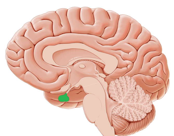 | |
Pituitary gland highlighted in green. click on image to enlarge
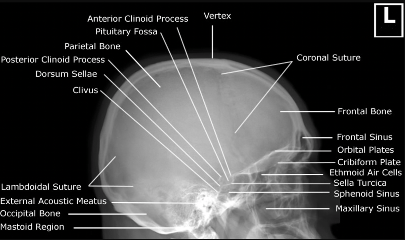 | |
Pituitary fossa and sella turcica on skull x-ray. click on image to enlarge
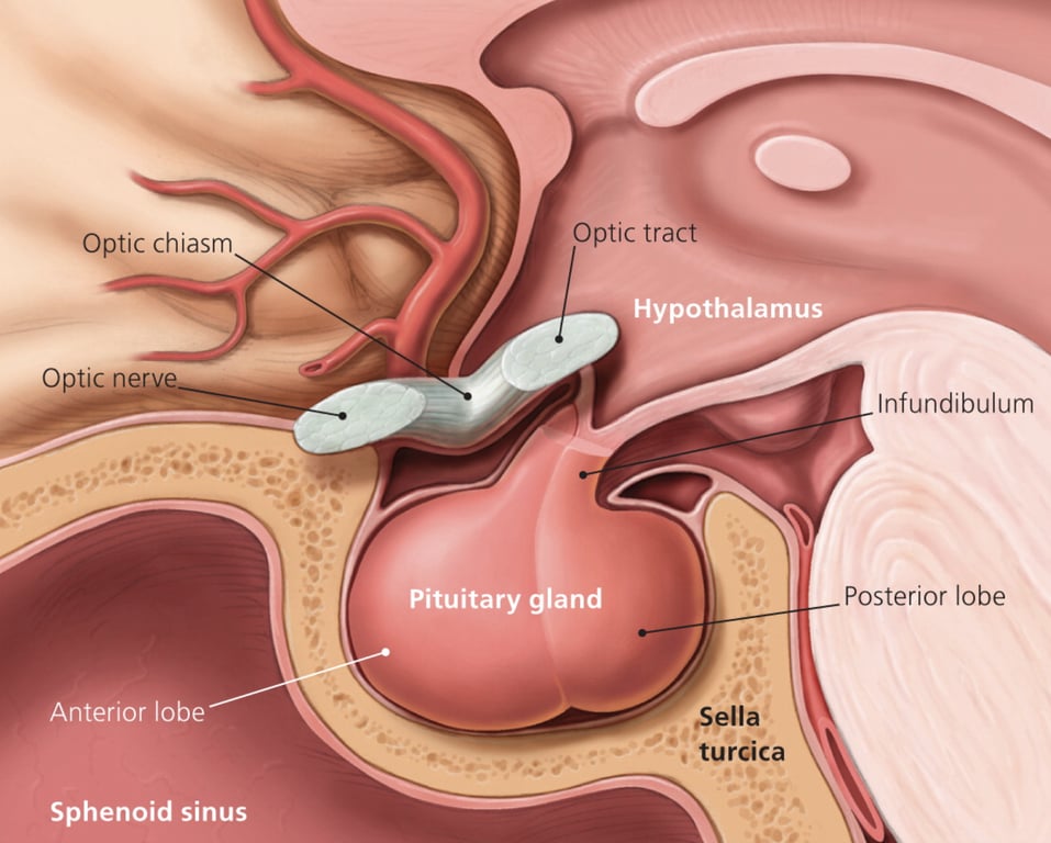 | |
Pituitary gland and relation to hypothalamus and optic chiasm. click on image to enlarge
Anterior and posterior pituitary
The anterior pituitary receives hormonal stimuli from the hypothalamus by way of the hypothalamo-pituitary portal system. The anterior pituitary is sometimes referred to as the adenohypophysis due to its non-neural nature.
The posterior pituitary is often considered an extension of the hypothalamus and is primarily composed of nervous tissue whose nuclei originate in the hypothalamus. The posterior pituitary is referred to as the neurohypophysis.
The posterior pituitary is actually an extension of the neurones of the paraventricular and supraoptic nuclei of the hypothalamus. The cell bodies of these regions rest in the hypothalamus, but their axons descend as the hypothalamic-hypophyseal tract within the infundibulum, and end in axon terminals that comprise the posterior pituitary
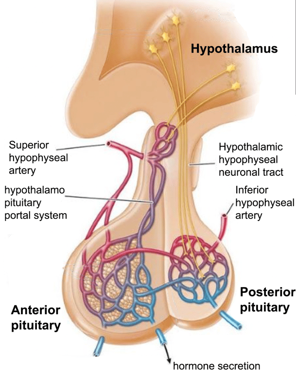 | |
Pituitary gland - anterior and posterior sections. click on image to enlarge
| Anterior pituitary | Posterior pituitary | |
|---|---|---|
| Derivation | Oral ectoderm | Neuroectoderm |
| Hormones secreted | ACTH TSH FSH LH GH Prolactin GH and prolactin are secreted by acidophilic cells ACTH, TSH, FSH & LH are secreted by basophilic cells These hormones are transported from hypothalamus to the anterior pituitary via the hypophyseal portal system | ADH Oxytocin |
ADH overview
Anti-diuretic hormone (ADH or vasopressin) is an important hormone in the control of water resorption and volume status. The synthesis of ADH occurs in the supraoptic and paraventricular nuclei in the hypothalamus. It is then transported to the posterior pituitary gland via the neurohypophysial capillaries.
ADH binding to V1 receptors causes vasoconstriction.
Increases in plasma concentration are detected by osmoreceptors in the hypothalamus which signal the posterior pituitary to release ADH. A fall in plasma volume is detected by barorecptors in the left atrium, aortic arch and carotid which signal the posterior pituitary to release ADH via the vagus nerve. Minor stimulants of ADH release include exercise, angiotensin II and some drugs such as nicotine.
A number of substance inhibit ADH release:
Anti-diuretic hormone (ADH or vasopressin) is an important hormone in the control of water resorption and volume status. The synthesis of ADH occurs in the supraoptic and paraventricular nuclei in the hypothalamus. It is then transported to the posterior pituitary gland via the neurohypophysial capillaries.
Mechanism of action
ADH binds to V2 receptors on renal principal cells in the distal convoluted tubule (DCT) and collecting ducts (CD). It stimulates the incorporation of aquaporin-2 channels into the apical membrane. The aquaporin channels allow increased water resorption and retention. As a result urine becomes more concentrated.ADH binding to V1 receptors causes vasoconstriction.
Control of secretion
ADH release is primarily controlled by plasma osmolality and plasma volume.Increases in plasma concentration are detected by osmoreceptors in the hypothalamus which signal the posterior pituitary to release ADH. A fall in plasma volume is detected by barorecptors in the left atrium, aortic arch and carotid which signal the posterior pituitary to release ADH via the vagus nerve. Minor stimulants of ADH release include exercise, angiotensin II and some drugs such as nicotine.
A number of substance inhibit ADH release:
- Alcohol
- Caffeine
- ANP (released when atria are stretched such as in fluid overload)
- Drugs e.g. steroids and ADH receptor antagonists