Eyelids
The eyelids are protective structures positioned in front of the eyes. The orbital septum (palpebral fascia) is the fibrous part of the eyelid and is an extension from the bony orbital margin that serves as a framework for the eyelids.
In the upper eyelid the septum blends with the tendon of the levator palpebrae superioris whilst in the lower lid the septum blends with the tarsal plate.
Innervation of the eyelids
Motor innervation:
Sensory Innervation upper eyelid:
Meibomian glands
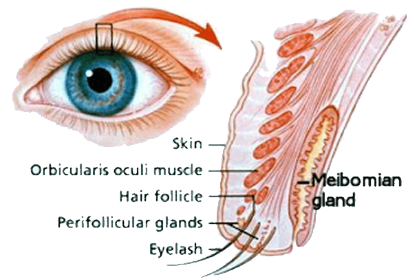
The meibomian (tarsal) glands are situated at the periphery of the eyelids. They secrete meibum which has a number of functions:
In the upper eyelid the septum blends with the tendon of the levator palpebrae superioris whilst in the lower lid the septum blends with the tarsal plate.
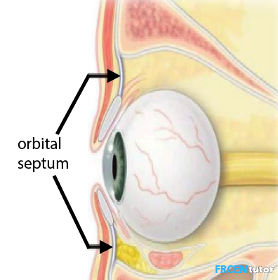 | |
The orbital septum (palpebral fascia) of the eyelids
Innervation of the eyelids
Motor innervation:
- Levator palpebrae superiorus (the main upper eyelid retractor) is innervated by oculomotor nerve (CN III)
- CN III also contributes fibres to lower eyelid retraction
- Sympathetic fibres innervate superior and inferior tarsal muscles contributing to lid retraction.
- Facial nerve innervates orbicularis oculi (the main eyelid protractor), frontalis, procerus and corrugator supercilii muscles supporting eyelid protraction.
Sensory Innervation upper eyelid:
- Supratrochlear
- Infratochlear
- Lacrimal
- Supraorbital
- Infratrochlear
- Infraorbital
- zygomaticofacial
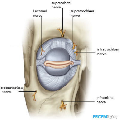 | |
Sensory innervation of the eyelids
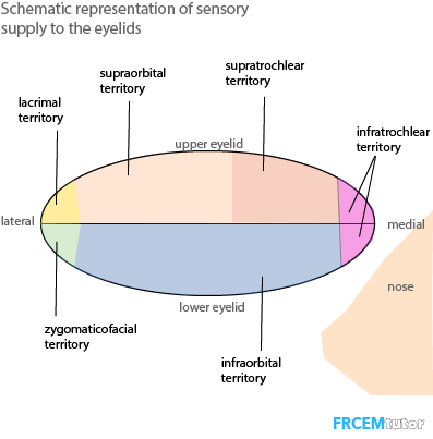 | |
Schematic view of sensory innervation of the eyelids
Meibomian glands
The meibomian (tarsal) glands are situated at the periphery of the eyelids. They secrete meibum which has a number of functions:
- Prevent tear film evaporation
- Prevent tear leakage onto cheek
- Forms tight seal when eyes closed
 | |
| Wikipedia | |