Uterus
Overview
The uterus is a hollow organ with thick muscular walls. The uterus can be divided anatomically into a fundus, body and cervix.Arterial supply
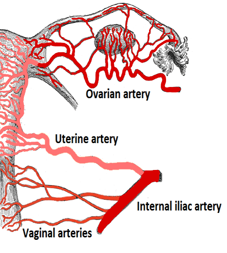
- The uterus is supplied by the uterine artery and the ovarian artery
- The uterine arteries are located in the Cardinal ligament (considered part of the Broad ligament)
The uterine arteries run within the Cardinal ligament. The Cardinal ligament is widely considered to be part of the Broad ligament (the inferior portion). if asked which structure the uterine arteries travel within and the Cardinal ligament is not an option then the Broad ligament is the answer.
Dimensions
- Uterus measures 8cm x 5cm
- The fallopian tube is appoximately 10cm in length.
Relations
| Anterior | Posterior | Lateral |
|---|---|---|
|
|
Lymph drainage
The uterine fundus drains to the para-aortic lymph nodes and superficial inguinal lymph nodes. The body and cervix of the uterus drain to the internal iliac lymph nodes.
Innervation of uterus and uterine tubes
- Innervation is from the inferior hypogastric plexus
- Parasympathetic supply originates from pelvic splanchnic nerves originating from S2-S4
- Sympathetic supply from fibres originating from T12-L2 spinal sections.
- Sensory afferent fibres mostly ascend through the inferior hypogastric plexus to enter the spinal cord via T10-T12 and L1 nerve fibres. From the cervix fibres travel along the pelvic splanchnic nerves and so pain related to cervical dilatation is referred to the sacral dermatomes.
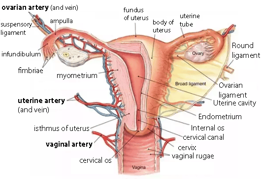 | |
Overview of uterine anatomy. Arterial supply highlighted
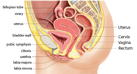 | |
Illustration showing lateral view of uterus and anatomical relations
 | |
| Wikipedia | |
Illustration highlighting arterial supply. Note the uterine arteries branch from the internal iliac whereas the ovarian arteries are direct branches of the aorta
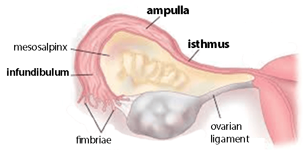 | |
Illustration showing uterine (fallopian) tubes and the main parts of the tube