Ureters
The ureters are muscular tubes extending from the kidneys to the bladder. They push urine along by peristaltic waves.
This knowledge allows the clinician to locate the likely location of stones on an X-ray KUB film.
Ureter innervation
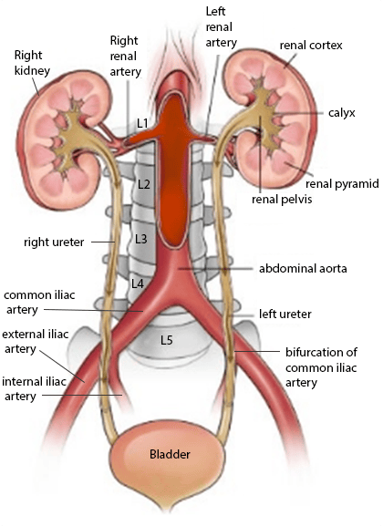 | |
Diagram illustrating the anatomy of the ureters. Note the ureters exit the hilum at L1 level. They then travel downward on the surface of the psoas muscle anterior to the tips of the transverse processes of the lumbar vertebrae. They enter the pelvis by crossing anterior to the bifurcation of the common iliac artery. In the pelvis they run down the lateral wall of the pelvis before turning obliquely at the level of the ischial spines to enter the posterolateral angles of the bladder
Ureter key anatomy
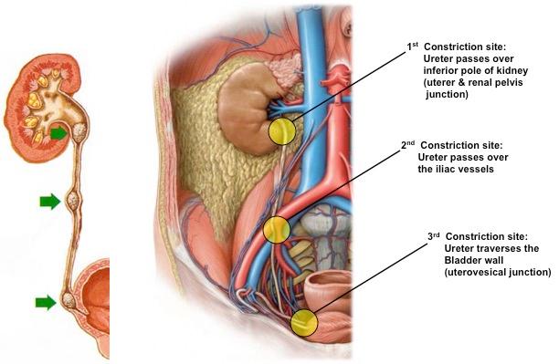
The ureters constrict at 3 places:- 1. Pelviureteric junction (PUJ): the joining of the renal pelvis to the abdominal part of the ureter
- 2. Where the ureters enter the pelvis in front of the sacro-iliac joint (at the bifurcation of the common iliacs)
- 3. Vesicoureteric junction (VUJ): where the ureters pierce the urinary bladder
This knowledge allows the clinician to locate the likely location of stones on an X-ray KUB film.
 | |
| Wikipedia | |
Ureter innervation
- Innervated via renal, testicular/ovarian and hypogastric plexuses
- Sensory fibres from the ureters enter the spinal cord at T11-L2 hence ureteric pain is referred to those dermatomes