Wound healing: Fracture healing
On this page
Phases of bone healing
1. Reactive (Inflammatory) phase (0–7 days)
- Vascular disruption leads to haematoma forms at fracture site.
- Platelets release cytokines and growth factors.
- Neutrophils and macrophages infiltrate, clear necrotic tissue, and release mediators.
- Haematoma & fibrin scaffold supports migration of reparative cells.
- Clinically: pain, swelling, and loss of function.
2. Reparative phase (1–6 weeks)
- Fibroblasts and chondroblasts migrate in and form fibrocartilaginous soft callus.
- Angiogenesis establishes granulation tissue and vascular supply.
- Osteoblasts deposit woven bone which converts soft callus into hard callus.
- Radiographic callus usually visible by 2–3 weeks.
- Clinical/radiological union:
- Children: often 3–6 weeks
- Adults: typically 6–12 weeks (long bones longer, up to ~3 months).
3. Remodelling phase (months–years)
- Woven bone replaced by lamellar bone.
- Osteoclasts resorb excess callus; medullary canal restored.
- Bone remodels along stress lines (Wolff’s law).
- Can take 1–2 years (occasionally longer).
- Final structure resembles original bone, with restoration of strength and shape.
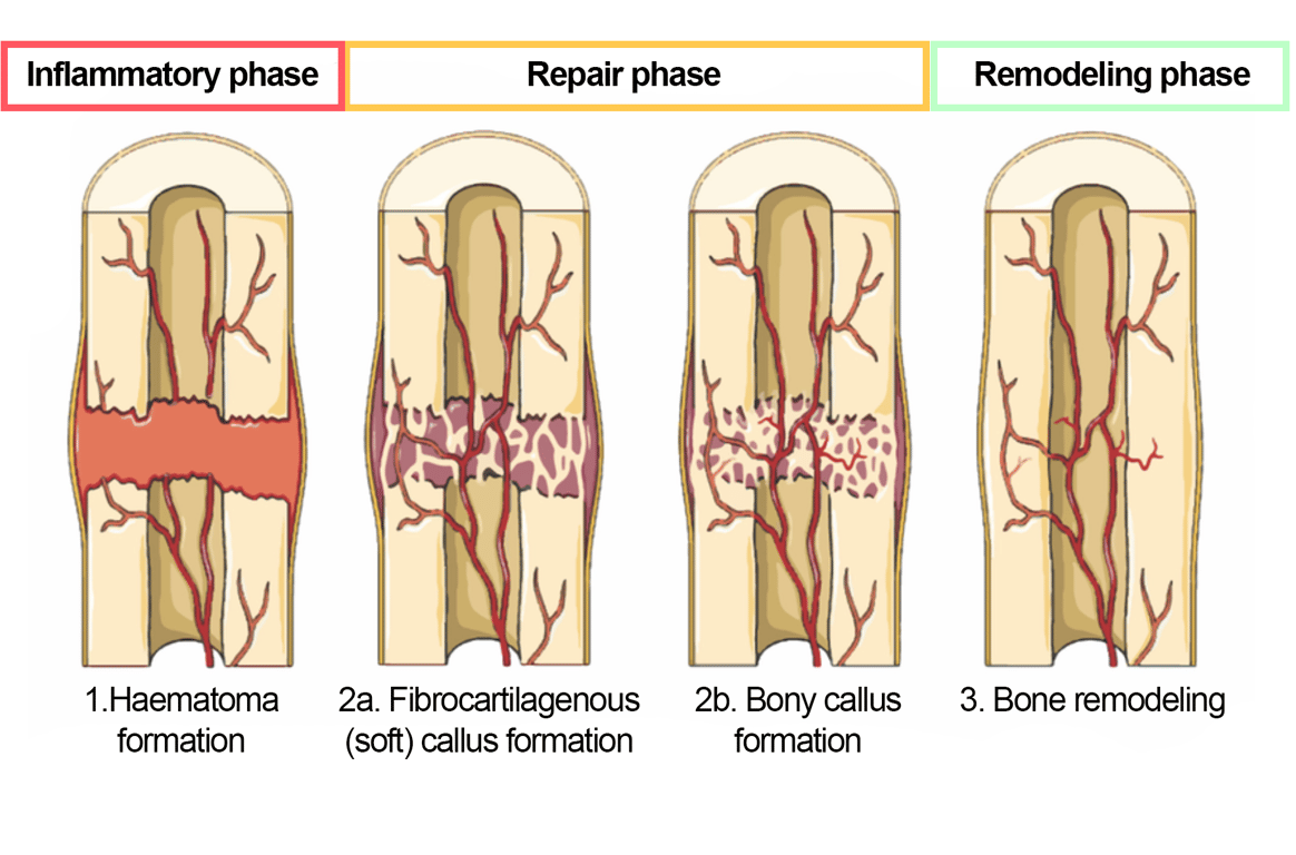 | |
Phases of fracture healing. Click on image to enlarge
Phase summary:
Haematoma → Soft callus → Hard callus → Remodelling
Haematoma → Soft callus → Hard callus → Remodelling
Complications of fracture healing
| Condition | Definition | Causes | Clinical features | Management |
|---|---|---|---|---|
| Delayed Union | Slower-than-expected healing of a fracture, usually taking longer than the normal healing period but eventually uniting. |
| Persistent pain, tenderness at the fracture site, minimal radiographic callus formation. | Optimize stability and blood supply; may involve bone stimulators or revision surgery in some cases. |
| Non-union | Failure of a fracture to unite after an extended period (typically 6–9 months), with no further progression toward healing. Types: Hypertrophic (adequate biology but insufficient stability); Atrophic (poor biology, often due to avascularity or severe soft tissue injury). |
| Persistent mobility or deformity at the fracture site, chronic pain, radiographs show persistent fracture line with absent bridging callus. | Surgical fixation, bone grafting, or use of bone stimulators depending on type and cause. |
| Malunion | Healing of a fracture in an incorrect anatomical position. |
| Deformity, altered limb length or angulation, functional impairment. | Corrective osteotomy may be required if function or appearance is compromised. |