Wound healing
On this page
Wound healing is a dynamic, overlapping process involving cellular and molecular mechanisms to restore tissue integrity. In the skin, it proceeds via four phases:
2. Inflammation
3. Proliferation
4. Remodeling / Maturation
Summary Table
Factors Affecting Healing
Complications of Healing
Wound healing in other tissues
The regenerative potential depends on whether tissue cells are:
- 1. Haemostasis
- 2. Inflammation
- 3. Proliferation
- 4. Remodeling.
Primary vs secondary intention
Primary intention:
Occurs when wound edges are closely opposed, e.g. a surgical incision. Minimal cell/tissue loss, clean and uninfected. Wound margins join by fibrin deposition, later replaced by collagen, with re-epithelialisation. Healing is rapid, scarring is minimal.
Secondary intention:
Occurs with extensive tissue loss, infection, or gaping wound edges. Defect fills with granulation tissue, epithelial regeneration covers the surface, and wound contraction occurs via myofibroblasts. Healing is slower and results in a larger scar.
Phases of Wound Healing
1. Hemostasis
- Immediate vasoconstriction, platelet adhesion/aggregation.
- Activation of the coagulation cascade leads to fibrin and fibronectin clot.
- Provides a waterproof barrier and provisional extracellular matrix.
- Platelets release PDGF and other mediators, attracting fibroblasts.
2. Inflammation
- Hours to days.
- Neutrophils dominate early (debris, microbes cleared).
- By day 3, macrophages replace neutrophils; secrete TGF-β, PDGF, VEGF, EGF.
- Keratinocytes at wound edges proliferate and migrate under the clot.
- Purpose: Debridement and initiation of repair.
3. Proliferation
- Days to weeks.
- Fibroblasts migrate in (2–5 days), proliferate, and secrete ECM (type III collagen, fibronectin).
- Angiogenesis (driven by VEGF): capillary buds → new vessels.
- Myofibroblasts contract the wound.
- Matrix metalloproteinases (MMPs) remodel ECM, enabling cellular/vascular migration.
4. Remodeling / Maturation
- Begins around day 5, can last months to more than 1 year.
- Type III collagen replaced by type I.
- Collagen fibers cross-link and align along stress lines.
- Vascular regression.
- Scar contracts (50–70% reduction in area).
- Tensile strength up to ~80% of normal by 12 weeks.
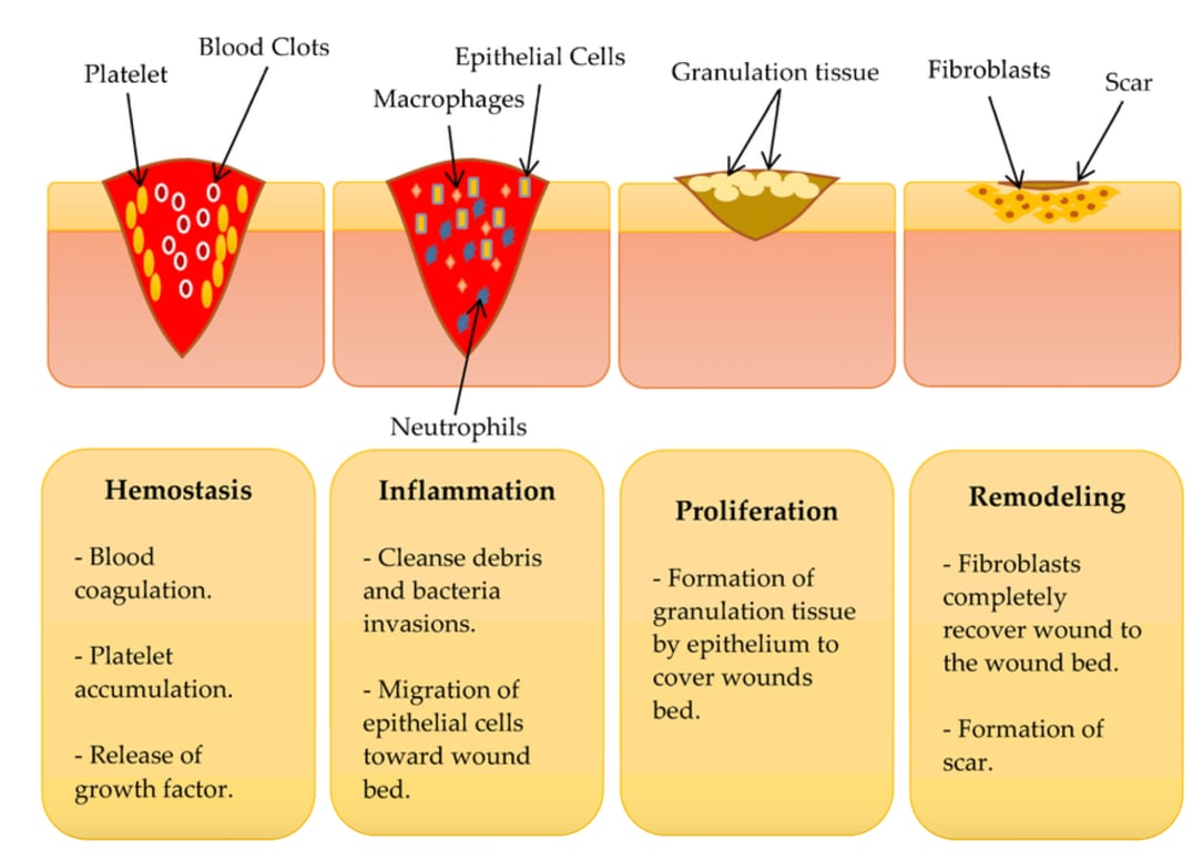 | |
Stages of wound healing. Click on image to enlarge
Summary Table
| Phase | Timing | Key Events & Mediators | Purpose |
|---|---|---|---|
| Hemostasis | Immediate – Hours | Platelet plug, fibrin/fibronectin clot, PDGF | Stop bleeding; provisional matrix |
| Inflammation | Days | Neutrophils → macrophages, cytokines (PDGF, TGF-β, VEGF, EGF) | Debridement, microbial defense, signaling |
| Proliferation | Days to Weeks | Fibroblasts (type III collagen, fibronectin), angiogenesis, keratinocytes, myofibroblasts, MMPs | Granulation tissue, contraction, epithelial cover |
| Remodeling | Weeks to >1 year | Type III → I collagen, alignment, vascular regression, scar contraction | Scar strengthening, structural restoration |
Cell types in wounds:
- Platelets – appear immediately (hemostasis and growth factor release)
- Neutrophils – infiltrate within the first 24 hours (peak early, phagocytosis of debris/microbes)
- Macrophages – appear at 24–48 hours (replace neutrophils, orchestrate inflammation and repair)
- Fibroblasts & myofibroblasts – appear from 2–4 days (ECM deposition, contraction)
- Endothelial cells – migrate/proliferate 3–5 days (angiogenesis)
Factors Affecting Healing
| Local | Systemic |
|---|---|
| Tissue hypoxia Infection Necrosis Foreign bodies Irradiation Repeated trauma Excessive wound edge tension | Age Diabetes mellitus Malnutrition (vitamin A, C, zinc) Smoking Obesity Glucocorticoid therapy Immunodeficiency |
Complications of Healing
- Scar contractures (e.g. burns, joints)
- Hypertrophic scars
- Keloids
- Failure to heal (chronic wounds, abscess, empyema)
- Wound dehiscence or hernia
- Traumatic neuroma.
- In fractures: delayed union, malunion, non-union.
Wound healing in other tissues
The regenerative potential depends on whether tissue cells are:
- Labile: continuously dividing (e.g. skin, GI epithelium, bone marrow). These tissues heal well.
- Stable: quiescent but can proliferate if stimulated (e.g. liver, kidney tubules, fibroblasts, endothelium). These tissues have limited regeneration.
- Permanent: non-dividing (e.g. neurons, cardiac myocytes). These tissues heal by scarring only.
| Category | Examples | Regenerative Capacity |
|---|---|---|
| Labile cells | Keratinocytes, intestinal epithelial cells, hematopoietic stem cells | High regenerative capacity; continual cell replacement |
| Stable cells | Hepatocytes, renal tubular epithelial cells, fibroblasts, vascular endothelial cells, tendon (tenocytes) | Limited regenerative capacity; quiescent at baseline but can proliferate following injury |
| Permanent cells | Neurons, cardiac myocytes, skeletal muscle cells (post-natal) | No regenerative capacity; injury leads to scarring or permanent loss of function |