Thalassaemias
On this page
Background
The thalassaemias are a heterogeneous group of inherited disorders of haemoglobin synthesis, caused by reduced or absent production of α or β-globin chains. The imbalance in globin chain synthesis leads to ineffective erythropoiesis, haemolysis, and variable anaemia.Normal haemoglobin structure
Normal adult haemoglobin (HbA) has four globin chains, two alpha and two beta (α₂β₂) and accounts for around 97% of circulating haemoglobin in healthy adults. Haemoglobin A2 (HbA₂) is comprised of two alpha and two delta chains (α₂δ₂ ) and typically accounts for 2–3% of total haemoglobin in healthy adults but is raised in β-thalassaemia trait. Fetal haemoglobin (HbF) accounts for <1% of haemoglobin in adults and is comprised of two alpha and two gamma chains (α₂γ₂).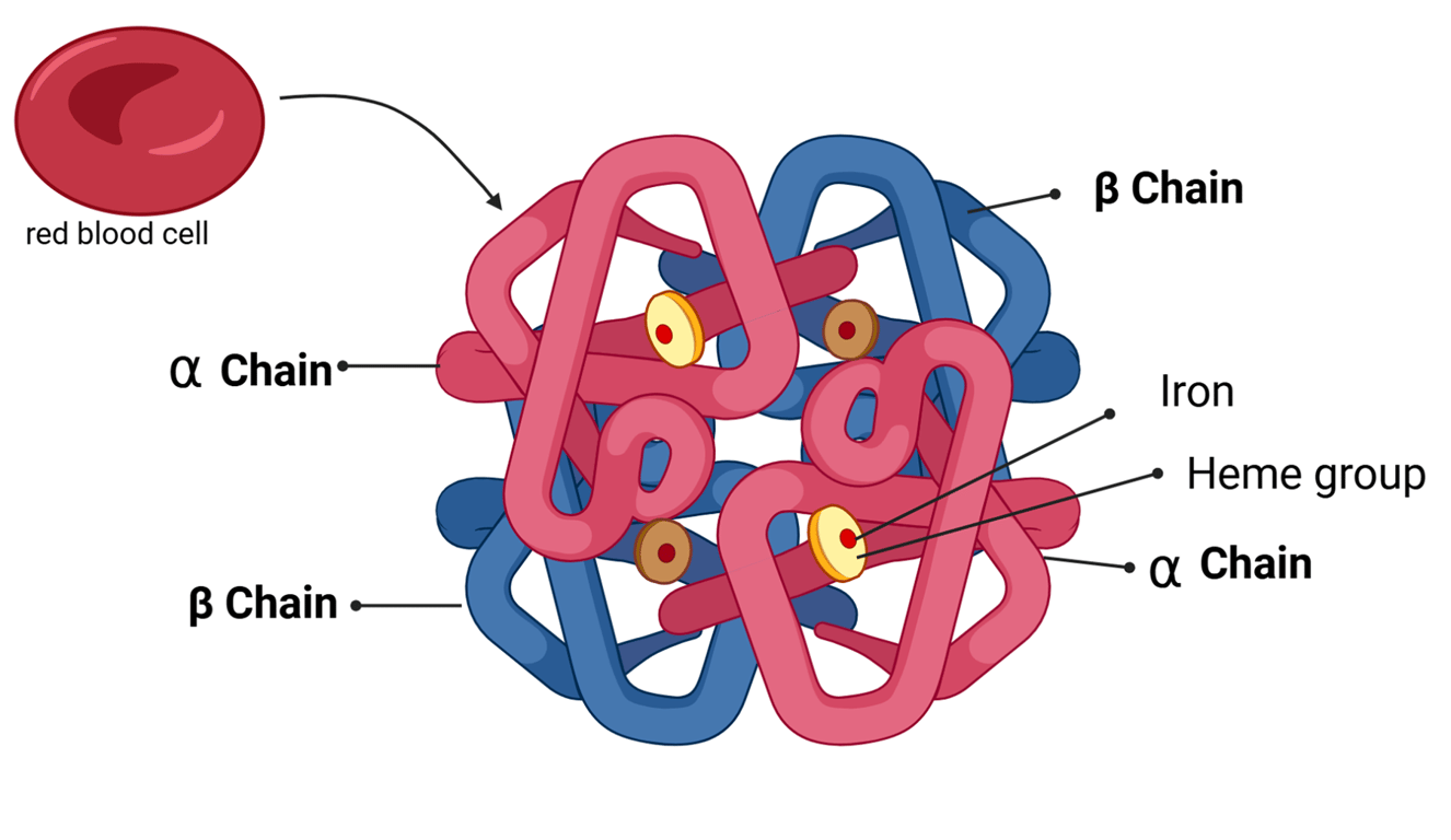 | |
Normal HbA structure. Click on image to enlarge
Epidemiology
β-thalassaemia: most prevalent in the Mediterranean, Middle East, South Asia, North Africa.
α-thalassaemia: more common in Southeast Asia, China, and Africa.
Genetics
α-thalassaemia: gene deletions/mutations across four α-globin alleles with spectrum from silent carrier, trait, HbH disease with haemolysis and splenomegaly, to Hb Bart’s hydrops fetalis.β-thalassemia is caused by point mutations (not deletions like α-thalassemia). These mutations affect the β-globin gene on chromosome 11. Point mutations may reduce or abolish β-globin production leading to phenotypes of trait (minor), intermedia and major. β-thalassaemia major typically presents in late infancy when HbF declines (failure to thrive, pallor, hepatosplenomegaly).
Inheritance:
autosomal recessive
Pathophysiology
A deficiency of one globin chain leads to an excess of the other. The unpaired chains precipitate in erythroblasts, causing membrane damage, ineffective erythropoiesis, apoptosis, and increased clearance by the reticuloendothelial system.Alpha-Thalassaemia
| Condition | Genetic Basis | Clinical Features |
|---|---|---|
| Silent carrier | 1 gene deleted | Normal or mild microcytosis |
| Trait | 2 genes deleted | Mild hypochromic, microcytic anaemia |
| HbH disease | 3 genes deleted | Moderate–severe anaemia, splenomegaly, haemolysis; HbH (β4 tetramers) seen |
| Hydrops foetalis | 4 genes deleted | Intrauterine death; no HbF or HbA production |
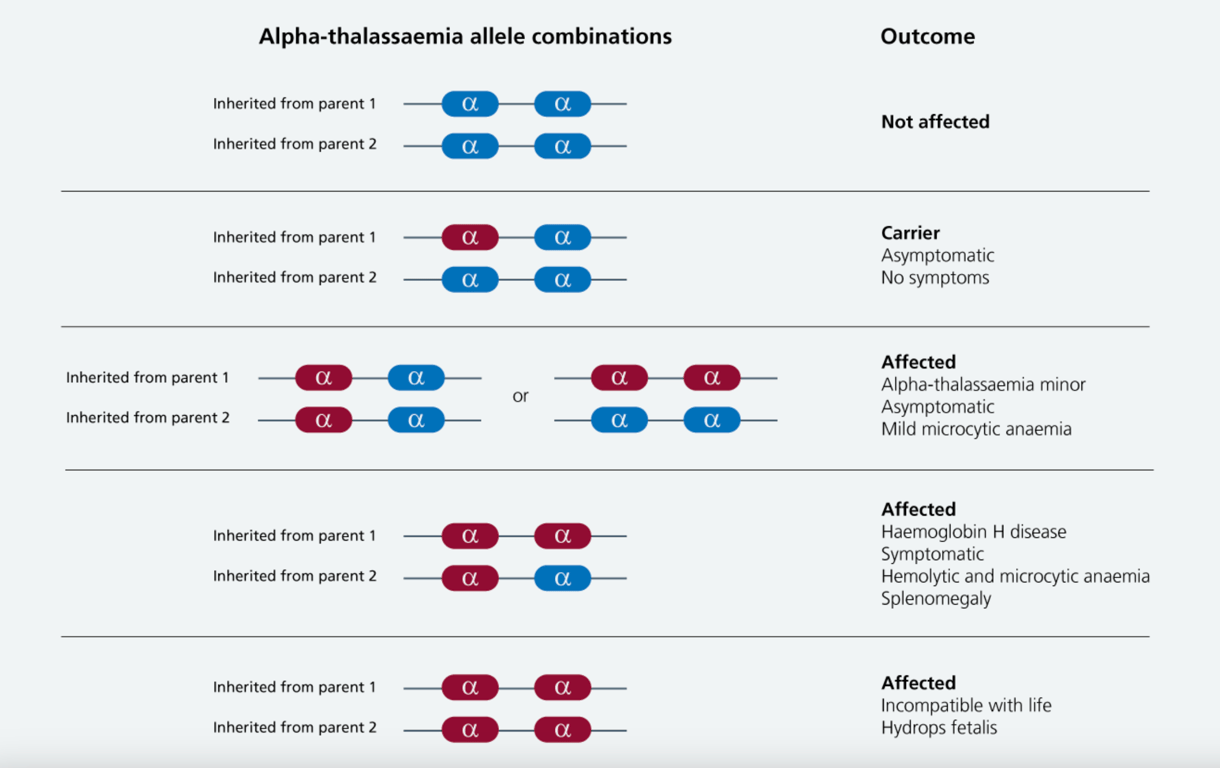 | |
Alpha-Thalassaemia inheritance visual summary. Click on image to enlarge
Beta-Thalassaemia
| Condition | Genetic Basis | Clinical Features |
|---|---|---|
| Trait (Minor) | 1 β-gene affected | Mild microcytic, hypochromic anaemia; raised RBC count; HbA₂ ↑; usually asymptomatic |
| Intermedia | Variable mutations | Moderate anaemia, variable transfusion requirement |
| Major (Cooley’s anaemia) | Both β-genes severely affected | Presents after 3–6 months when HbF levels decline; severe anaemia, failure to thrive, jaundice, hepatosplenomegaly, bone marrow expansion leads to bone changes (thalassaemic facies, bossing, 'hair-on-end' skull x-ray); iron overload from transfusions/absorption |
Investigations
- FBC: microcytic, hypochromic anaemia.
- Blood film: target cells, nucleated red cells; in β-major, erythroblasts and myelocytes.
- Bone marrow: erythroid hyperplasia.
- Hb electrophoresis: β-trait: HbA₂ >3.5%, β-major: ↑HbF, little or no HbA, α-thal diagnosis by gene deletion analysis.
Complications
- Anaemia and ineffective erythropoiesis.
- Extramedullary haematopoiesis (splenomegaly, bone deformities).
- Iron overload (transfusion-related + increased absorption).
- Growth delay, endocrine dysfunction, cardiomyopathy.
Management
- β-major: regular transfusions, iron chelation (desferrioxamine, deferasirox), splenectomy in selected cases, curative stem cell transplant.
- Genetic counselling and antenatal screening important for prevention.
Management (principles)
Public health & screening
Carrier screening, partner testing, and antenatal/newborn programmes reduce incidence and guide reproductive choices; counselling is essential
- Transfusion programmes for transfusion-dependent thalassaemia (TDT) to suppress ineffective erythropoiesis and maintain growth/quality of life.
- Iron chelation (e.g., deferasirox, deferiprone, deferoxamine) guided by ferritin and MRI liver/heart iron.
- Splenectomy for hypersplenism (selected cases), with vaccination and prophylaxis.
- Endocrine, hepatic and cardiac monitoring for iron overload complications.
- Curative options: haematopoietic stem cell transplantation in suitable candidates; gene therapy is emerging (e.g., betibeglogene autotemcel) in specialised centres.
Public health & screening
Carrier screening, partner testing, and antenatal/newborn programmes reduce incidence and guide reproductive choices; counselling is essential