Acquired coagulation disorders
Acquired coagulation disorders develop after birth secondary to systemic disease, drugs, or autoantibodies. They are more common than inherited disorders and often involve multiple factor deficiencies.
Key acquired coagulation disorders include:
These are considered in more detail below:
Vitamin K deficiency
Causes:
Reduced activity of vitamin K–dependent factors (II, VII, IX, X, protein C, protein S)
Labs:
Liver disease-associated coagulopathy
Pathophysiology:
Disseminated intravascular coagulation (DIC)
Pathogenesis:
Treat underlying cause, supportive transfusion (FFP, platelets, cryoprecipitate); anticoagulation in select thrombotic cases
Autoantibody inhibition of coagulation
Specific inhibitors:
Miscellaneous causes
Summary table
Key acquired coagulation disorders include:
- Vitamin K deficiency
- Liver disease associated coagulopathy
- Disseminated intravascular coagulation (DIC)
- Autoantibody inhibition of coagulation
- Other miscellaneous causes e.g. transfusion syndrome
These are considered in more detail below:
Vitamin K deficiency
Causes:
- Haemorrhagic disease of the newborn
- Biliary obstruction (fat malabsorption)
- Malnutrition/malabsorption (e.g. pancreatic, bowel disease)
- Vitamin K antagonist therapy (warfarin)
- Prolonged antibiotic use (↓ gut flora)
Reduced activity of vitamin K–dependent factors (II, VII, IX, X, protein C, protein S)
Labs:
- Prolonged PT (early)
- Prolonged aPTT in severe cases
- Normal platelet count
- Mucocutaneous bleeding
- post-surgical haemorrhage
- neonatal bleeding
Vitamin K is a fat-soluble vitamin, obtained from:
- Dietary sources (especially green leafy vegetables)
- Bacterial synthesis in the gut (colon flora)
- Newborns due to low placental transfer, immature liver, low vitamin K stores, and lack of gut flora (known as haemorrhagic disease of the newborn)
- Later life due to malnutrition, fat malabsorption (biliary obstruction, pancreatic disease), prolonged antibiotics, or vitamin K antagonist therapy (warfarin)
Liver disease-associated coagulopathy
Pathophysiology:
- Decreased synthesis of clotting factors (all except factor VIII)
- Decreased fibrinogen in severe hepatocellular failure
- Dysfibrinogenemia (abnormal fibrinogen function)
- Increased Plasminogen activator → enhanced fibrinolysis
- Decreased Thrombopoietin → thrombocytopenia
- Hypersplenism (portal hypertension) → splenic sequestration of platelets
- Biliary obstruction → vitamin K malabsorption
- DIC may complicate severe disease
- Net effect: Can be prothrombotic or haemorrhagic
- Prolonged PT
- Prolonged aPTT
- Low fibrinogen (in severe cases)
- Thrombocytopenia
Disseminated intravascular coagulation (DIC)
Pathogenesis:
- Widespread activation of coagulation → excess thrombin → fibrin deposition and platelet consumption
- Simultaneous activation of fibrinolysis → fibrin degradation products (FDPs) interfere with fibrin polymerisation
- Net result: consumptive coagulopathy (bleeding + thrombosis)
- Infection: Gram-negative sepsis, meningococcal sepsis, malaria
- Malignancy: acute leukaemia, adenocarcinoma
- Obstetric: amniotic fluid embolism, placental abruption, eclampsia
- Hypersensitivity: anaphylaxis, incompatible transfusion
- Tissue damage: trauma, burns, surgery
- Vascular: leaking prosthetic valves, bypass surgery, aneurysms
- Misc: liver failure, snake bites, pancreatitis, hypothermia, massive haemorrhage
- Bleeding: from venepuncture sites, wounds, GI tract, mucosa, urogenital tract, obstetric bleeding
- Thrombosis: skin necrosis, renal failure, gangrene, cerebral ischaemia
- Decreased platelets
- Decreased fibrinogen
- Increased FDPs / D-dimer
- Prolonged PT, aPTT, TT
- Evidence of haemolysis
Treat underlying cause, supportive transfusion (FFP, platelets, cryoprecipitate); anticoagulation in select thrombotic cases
Autoantibody inhibition of coagulation
Specific inhibitors:
- Acquired haemophilia A (autoantibodies to factor VIII) → severe bleeding
- Lupus anticoagulant / antiphospholipid antibodies (often prothrombotic despite prolonged aPTT)
Miscellaneous causes
- Diseases with M-protein production (e.g. multiple myeloma) → interference with coagulation
- L-asparaginase therapy (decreased protein synthesis)
- Anticoagulant therapies (Heparin, defibrinating agents e.g. snake venom, thrombolytic therapy)
- Massive transfusion syndrome → dilutional coagulopathy (loss of clotting factors + platelets)
- Uremia → platelet dysfunction (normal PT/aPTT, prolonged bleeding time)
Summary table
| Cause | Pathophysiology | Labs | Clinical Features |
|---|---|---|---|
| Vitamin K deficiency (newborn, biliary obstruction, malabsorption, warfarin) | ↓ vitamin K–dependent factors (II, VII, IX, X, protein C & S) | Prolonged PT (early), prolonged aPTT if severe, normal platelets | Mucocutaneous bleeding, surgical/postpartum haemorrhage, neonatal bleeding |
| Liver disease | ↓ synthesis of clotting factors (except VIII), ↓ factor V & fibrinogen, dysfibrinogenemia, ↑ fibrinolysis, ↓ thrombopoietin, hypersplenism | Prolonged PT & aPTT, ↓ fibrinogen, thrombocytopenia | Bruising, mucosal bleeding, variceal haemorrhage, paradoxical thrombosis |
| DIC | Widespread thrombin activation → fibrin deposition, platelet & factor consumption, ↑ fibrinolysis → FDPs | ↓ Platelets, ↓ fibrinogen, ↑ D-dimer, prolonged PT, aPTT, TT, evidence of haemolysis | Bleeding (venepuncture, mucosa, GI, obstetric) + thrombosis (skin necrosis, renal failure, CNS ischaemia) |
| Acquired hemophilia (specific inhibitor) | Autoantibodies against factor VIII | Isolated prolonged aPTT, normal PT, normal platelets | Severe spontaneous bleeding (soft tissue, mucosa, GI, GU) |
| Lupus anticoagulant (non-specific inhibitor) | Antiphospholipid antibodies interfere with phospholipid-dependent assays | Prolonged aPTT (not corrected on mixing), PT normal, platelets normal | Often prothrombotic (DVT, PE, miscarriage) despite prolonged aPTT |
| Miscellaneous (M-protein, L-asparaginase, heparin, thrombolytics, massive transfusion, uremia) | Protein interference, ↓ synthesis, drug effects, dilution, platelet dysfunction | Variable: heparin → prolonged aPTT, thrombolytics → low fibrinogen, massive transfusion → low platelets & factors, uremia → normal PT/aPTT but prolonged bleeding time | Bleeding tendency, sometimes thrombosis (esp. heparin-induced thrombocytopenia) |
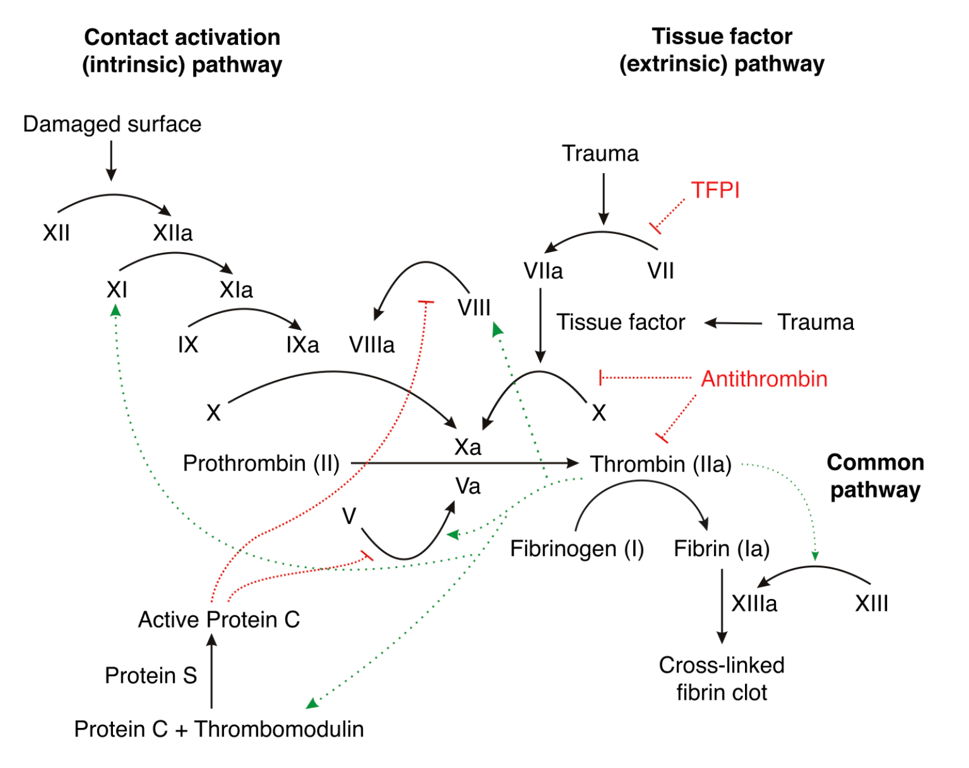 | |
Coagulation pathway. Click on image to enlarge