Calcium homeostasis: PTH, Vitamin D & Calcitonin
Calcium is an important ion that plays a key role in a number of cellular functions including:
As calcium is a trigger to many crucial cellular events it is important for free Ca2+ ion levels to be carefully controlled. Plasma calcium levels are controlled by the co-ordinated action of parathyroid hormone (PTH), activated vitamin D and calcitonin.
Chief cells have membrane bound receptor proteins that bind plasma Ca2+ ions. When plasma Ca2+ ion levels rise receptor binding decreases intracellular cAMP levels which inhibits PTH release. In contrast low Ca2+ ion levels result in rising cAMP and PTH release.
PTH secretion may also be stimulated by:
Actions of PTH
PTH increases phosphate excretion by inhibiting reabsorption in the proximal tubule of the nephron.
PTH also inhibits bicarbonate reabsorption promoting a more acidic pH. Acidosis reduces calcium binding to albumin increasing free calcium ion levels.
The Ca2+-ATPase pump on the basolateral membrane is also activated by active vitamin D.
A Na+-Ca2+ antiporter on the basolateral membrane also assists in driving calcium into the interstitium (see the image below).
Stimulates hydroxylation of vitamin D in the kidneys to produce activated vitamin D (activated vitamin D is known as 1,25-dihydroxycholecalciferol or calcitriol)
Calcitonin
Calcitonin is released from C-cells (parafollicular cells) of the thyroid gland in response to high plasma Ca2+ levels.
Calcitonin may simplistically be thought of as antagonistic to the actions of PTH and vitamin D on plasma calcium levels.
Calcitonin acts at multiple sites to achieve this:
Vitamin D
Vitamin D refers to a group of fat soluble steroid molecules. In the human diet vitamin D is typically ingested in two forms:
Vitamin D3 may also be synthesised in the lower layers of the epidermis from cholesterol via a UV light driven reaction.
Vitamin D from the diet, or from skin synthesis, is biologically inactive. It is activated by two enzymatic hydroxylation steps, the first in the liver and the second in the kidneys. The second hydroxylation in the renal tubule is under the influence of PTH and is the slowest and therefore rate limiting step. Vitamin D is considered to be a hormone.
The second vitamin D hydroxylation which leads to the formation of active vitamin D (calcitriol or 1,25 Dihydroxyvitamin D3) occurs in the proximal tubules. Activated vitamin D3 then acts on the distal renal tubule cells to increase renal tubular reabsorption of calcium via activation of basolateral Ca2+ ATPase pumps.
Activated vitamin D promotes calcium and phosphate resorption in the intestines and promotes osteoclast activity to release calcium and phosphate from bone. Activated vitamin D also suppresses PTH secretion.
Vitamin D and its effect on bone may seem confusing as increasing osteoclast activity would have the expected effect of decreasing bone mass, after all PTH increases osteoclast activity and hyperparathyroidism is associated with osteoporosis.
Studies show that vitamin D has the opposite effect however and improves bone mineral density. The leading hypothesis is that its actions provide optimum serum mineral levels for new bone mineralisation to occur as both calcium and phosphorus are key minerals for bone mineralization.
At first glance it may seem that PTH and vitamin D have similar roles but PTH acts to maintain serum calcium levels within a tight range at the expense of maintaining adequate phosphate. Vitamin D promotes both calcium and phosphate resorption & inhibits PTH.
- Muscle contraction
- Neurotransmitter release
- Hormone release
- Secretion from exocrine glands
- Activation of enzymes
As calcium is a trigger to many crucial cellular events it is important for free Ca2+ ion levels to be carefully controlled. Plasma calcium levels are controlled by the co-ordinated action of parathyroid hormone (PTH), activated vitamin D and calcitonin.
99% of body calcium is stored in the skeleton as hydroxyapatite. The remaining 1% is predominantly found in extracellular fluid. Within cells the majority of free calcium is stored in the endoplasmic reticulum or mitochondria.
If extracellular Ca2+ levels drop too low it triggers the spontaneous opening of Na+ channels causing involuntary muscle contractions. This clinical sign of hypocalcaemia is termed hypocalcaemic tetany.
If extracellular Ca2+ levels drop too low it triggers the spontaneous opening of Na+ channels causing involuntary muscle contractions. This clinical sign of hypocalcaemia is termed hypocalcaemic tetany.
Parathyroid hormone
Parathyroid hormone (PTH) is the major controller of plasma calcium. PTH is primarily secreted by chief cells of the parathyroid glands.The net effect of PTH is to increase plasma calcium levels and decrease plasma phosphate levels
Chief cells have membrane bound receptor proteins that bind plasma Ca2+ ions. When plasma Ca2+ ion levels rise receptor binding decreases intracellular cAMP levels which inhibits PTH release. In contrast low Ca2+ ion levels result in rising cAMP and PTH release.
PTH secretion may also be stimulated by:
- Mild hypomagnesaemia (but note severe hypomagnesaemia paradoxically inhibits PTH release)
- Increase in serum phosphate (increased phosphate causes it to complex with serum calcium, forming calcium phosphate, which lowers free calcium available to bind to receptors).
- Adrenaline & histamine
Actions of PTH
| Site | Action |
|---|---|
| Bone | Increased osteocytic osteolysis Increased osteoclast activity The above releases Ca2+ (and phosphate) ions by bone breakdown (resorption) |
| Kidney | Increases Ca2+ reabsorption (see below) Stimulates production of activated vitamin D |
| Intestine | Indirectly enhances absorption of Ca2+ (and phosphate) ions PTH converts 25-hydroxy vitamin D into 1,25-dihydroxy vitamin D (calcitriol) which promotes calcium reabsorption from the intestine |
PTH and its action on the kidney
PTH activates Ca2+ entry channels on the apical membrane and the Ca2+-ATPase pump on the basolateral membrane. Calcium flows into the tubule cells via the Ca2+ entry channels where binding proteins prevent free ionised calcium levels from rising too high.PTH increases phosphate excretion by inhibiting reabsorption in the proximal tubule of the nephron.
PTH also inhibits bicarbonate reabsorption promoting a more acidic pH. Acidosis reduces calcium binding to albumin increasing free calcium ion levels.
The Ca2+-ATPase pump on the basolateral membrane is also activated by active vitamin D.
A Na+-Ca2+ antiporter on the basolateral membrane also assists in driving calcium into the interstitium (see the image below).
Stimulates hydroxylation of vitamin D in the kidneys to produce activated vitamin D (activated vitamin D is known as 1,25-dihydroxycholecalciferol or calcitriol)
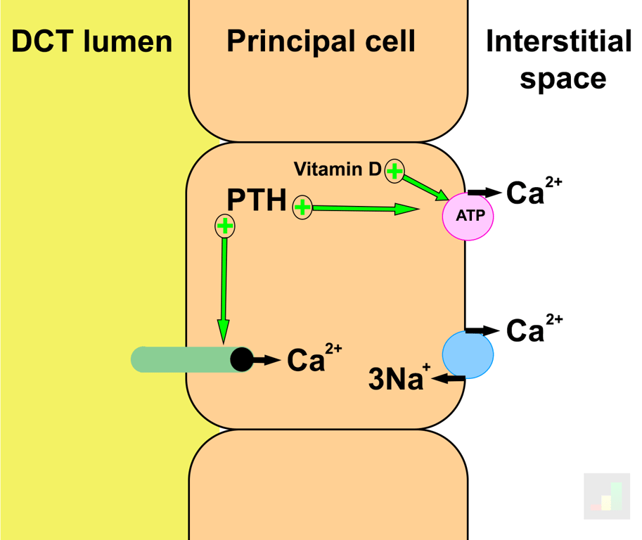 | |
Renal handling of calcium in the distal tubule. Click on image to enlarge
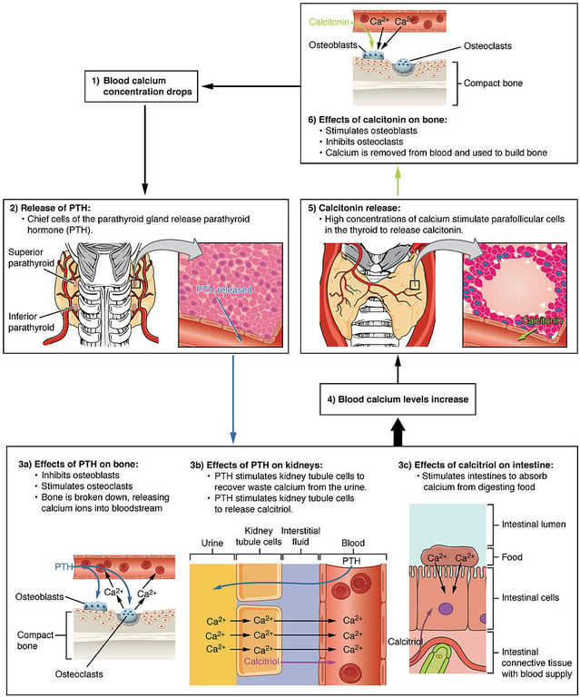 | |
Calcium homeostatsis. The roles of PTH and calcitonin. Click on image to enlarge
Calcitonin
Calcitonin is released from C-cells (parafollicular cells) of the thyroid gland in response to high plasma Ca2+ levels.
Calcitonin may simplistically be thought of as antagonistic to the actions of PTH and vitamin D on plasma calcium levels.
Calcitonin acts to lower plasma calcium levels.
Calcitonin acts at multiple sites to achieve this:
- In bone calcitonin inhibits bone breakdown and resorption of Ca2+ by osteoclasts. It is also thought to promote osteoblast utilisation of Ca2+ to form new bone.
- In the kidneys calcitonin inhibits Ca2+ and PO43- resorption by renal tubular cells which promotes their excretion.
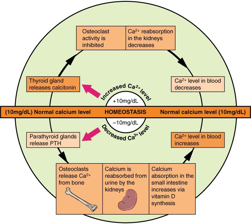 | |
Calcium homeostasis. Click on image to enlarge
Vitamin D
Vitamin D refers to a group of fat soluble steroid molecules. In the human diet vitamin D is typically ingested in two forms:
- Vitamin D2 (ergocalciferol) derived form plants and yeasts
- Vitamin D3 (cholecalciferol) derived from animal products especially dairy
Vitamin D3 may also be synthesised in the lower layers of the epidermis from cholesterol via a UV light driven reaction.
Vitamin D from the diet, or from skin synthesis, is biologically inactive. It is activated by two enzymatic hydroxylation steps, the first in the liver and the second in the kidneys. The second hydroxylation in the renal tubule is under the influence of PTH and is the slowest and therefore rate limiting step. Vitamin D is considered to be a hormone.
The second vitamin D hydroxylation which leads to the formation of active vitamin D (calcitriol or 1,25 Dihydroxyvitamin D3) occurs in the proximal tubules. Activated vitamin D3 then acts on the distal renal tubule cells to increase renal tubular reabsorption of calcium via activation of basolateral Ca2+ ATPase pumps.
Activated vitamin D promotes calcium and phosphate resorption in the intestines and promotes osteoclast activity to release calcium and phosphate from bone. Activated vitamin D also suppresses PTH secretion.
Activated vitamin D actions:
| Site | Action |
|---|---|
| Intestine | Increases calcium and phosphate absorption in the small intestine |
| Kidney | Increases renal tubular reabsorption of calcium via activation of basolateral Ca2+ ATPase Increases renal tubular reabsorption of phosphate |
| Bone | Stimulates release of calcium from bone (Indirectly activates osteoclasts via stimulation of osteoblasts to release RANKL) |
| Parathyroid gland | Inhibits PTH secretion |
The net effect of activated vitamin D is to increase plasma calcium and phosphate levels
Vitamin D and its effect on bone may seem confusing as increasing osteoclast activity would have the expected effect of decreasing bone mass, after all PTH increases osteoclast activity and hyperparathyroidism is associated with osteoporosis.
Studies show that vitamin D has the opposite effect however and improves bone mineral density. The leading hypothesis is that its actions provide optimum serum mineral levels for new bone mineralisation to occur as both calcium and phosphorus are key minerals for bone mineralization.
At first glance it may seem that PTH and vitamin D have similar roles but PTH acts to maintain serum calcium levels within a tight range at the expense of maintaining adequate phosphate. Vitamin D promotes both calcium and phosphate resorption & inhibits PTH.