Retroperitoneal structures
The peritoneum is a double layered serous membrane. It is composed of visceral and parietal layers with serous fluid in a small cavity between the two layers that allows the layers to glide over one another.
Parietal peritoneum lines the walls of the abdomen and pelvis whereas visceral peritoneum covers the organs. Retroperitoneal organs have peritoneum covering them anteriorly but not posteriorly. They are considered less vulnerable to deceleration injuries than intraperitoneal organs.
A useful pneumonic to remember the retroperitoneal organs is:
SAD PUCKER see below:
S = Suprarenal glands (adrenal glands)
A = Aorta/IVC
D = Duodenum (2nd, 3rd and part of 4th segment)
P = Pancreas (tail is intraperitoneal)
U = Ureters
C = Colon (ascending and descending colons only - transverse and sigmoid have mesentaries)
K = Kidneys
E = Esophagus
R = Rectum
NOTE: The retroperitoneal organs are less likely to be injured in deceleration. In deceleration injuries shear forces put organs with free mesenteries at high risk. Mesenteric tears and hepatic tears are common. Retroperitoneal injury can occur through injury to their support structures. Most commonly this is seen with shearing of the renal arteries with resultant renal injury secondary to this.
Parietal peritoneum lines the walls of the abdomen and pelvis whereas visceral peritoneum covers the organs. Retroperitoneal organs have peritoneum covering them anteriorly but not posteriorly. They are considered less vulnerable to deceleration injuries than intraperitoneal organs.
Retroperitoneal organs and structures
A useful pneumonic to remember the retroperitoneal organs is:
SAD PUCKER see below:
S = Suprarenal glands (adrenal glands)
A = Aorta/IVC
D = Duodenum (2nd, 3rd and part of 4th segment)
P = Pancreas (tail is intraperitoneal)
U = Ureters
C = Colon (ascending and descending colons only - transverse and sigmoid have mesentaries)
K = Kidneys
E = Esophagus
R = Rectum
Retroperitoneal vs intraperitoneal
| Retroperitoneal structures | Intraperitoneal structures |
|---|---|
|
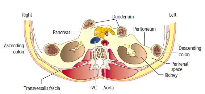 | |
Image illustrating the retroperitoneal structures in yellow
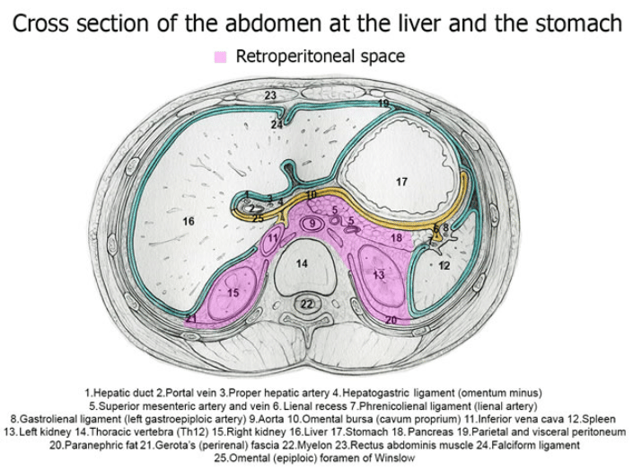 | |
More detailed overview of the retroperitoneal structures. Retroperitoneal space is highlighted in purple
NOTE: The retroperitoneal organs are less likely to be injured in deceleration. In deceleration injuries shear forces put organs with free mesenteries at high risk. Mesenteric tears and hepatic tears are common. Retroperitoneal injury can occur through injury to their support structures. Most commonly this is seen with shearing of the renal arteries with resultant renal injury secondary to this.