Rectus sheath
The rectus abdominis are contained within the rectus sheath above the arcuate line. Below the arcuate line the rectus sheath is anterior to the rectus abdominis.
The rectus sheath is formed by blending of the aponeurosis of the 3 main abdominal muscles ie the external oblique, the internal oblique and transversus.
As can be seen from the diagrams the sheath splits to encompass the rectus abdominis above the arcuate line.
The sheath contains the following 3 structures:
- Inferior epigastric arteries
- Superior epigastric arteries
- Posterior intercostal nerves
The diagrams show how the rectus sheath forms and how it changes above and below the arcuate line (horizontal line usually just below umbilicus that represents where the posterior layer of the rectus sheath ends)
Diagram illustrating the rectus sheath above and below the arcuate line
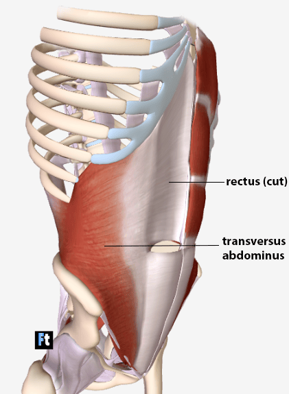
Diagram illustrating transversus abdominus anterior and lateral views
The rectus sheath is formed by blending of the aponeurosis of the 3 main abdominal muscles ie the external oblique, the internal oblique and transversus.
As can be seen from the diagrams the sheath splits to encompass the rectus abdominis above the arcuate line.
The sheath contains the following 3 structures:
- Inferior epigastric arteries
- Superior epigastric arteries
- Posterior intercostal nerves
The diagrams show how the rectus sheath forms and how it changes above and below the arcuate line (horizontal line usually just below umbilicus that represents where the posterior layer of the rectus sheath ends)
Diagram illustrating the rectus sheath above and below the arcuate line
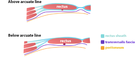 | |
Diagram illustrating transversus abdominus anterior and lateral views
 | |
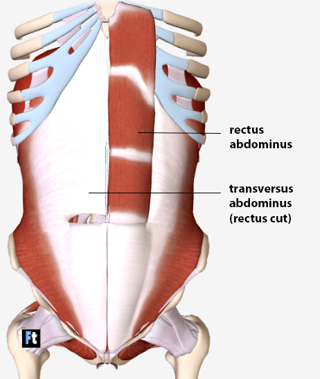 | |
Note the two most superficial layers, the external and internal oblique, have been removed. This shows the transversus abdominus muscle running behind the rectus superiorly (above the arcuate line) but the rectus pierces the transversus and lies posterior to it in the lower 1/4 of the abdominal wall i.e. below the arcuate line