Respiratory physiology: Control of breathing
On this page
Control of ventilation
The control of breathing is essential to facilitate respiration and maintain satisfactory levels of oxygen, carbon dioxide and blood pH.Breathing is primarily controlled by the respiratory centres of the autonomic nervous system. The majority of our breathing is under involuntary control.
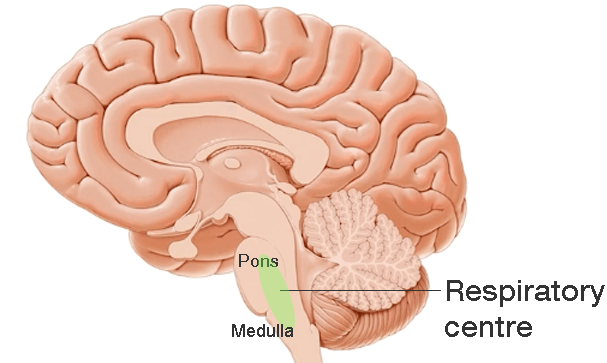
The respiratory centres of the brain are collections of neurones in the pons and medulla of the brainstem. They receive sensory information from chemoreceptors and mechanoreceptors.
In the notes below we will consider:
- Chemoreceptors as the primary sensory input controlling ventilation
- The respiratory centres of the brain that process sensory input and co-ordinate ventilation
- Voluntary control of ventilation
Chemical control of respiration: Central & peripheral chemoreceptors
There are two types of chemoreceptors involved in controlling ventilation:
- Central chemoreceptors
- Peripheral chemoreceptors
| Receptor | Location | Role |
|---|---|---|
| Central chemoreceptor | Medulla | Respond to changes in:
|
| Peripheral chemoreceptors | Carotid bodies Aortic bodies | Respond to changes in:
|
Central chemoreceptors
Central chemoreceptors are located in the medulla only a few microns from the medullary respiratory groups.
The central chemoreceptor is the major determinant of respiratory response. It responds to changes in pH. Note it does not directly measure pCO2 but responds to changes in pH as a result of CO2 changes. It accounts for approx 80% of respiratory response.
CO2 easily diffuses across the blood brain barrier and makes the CSF more acidic. The central chemoreceptor will detect this and send signals to the respiratory centre to effect a rise in respiratory rate to 'blow off' CO2.
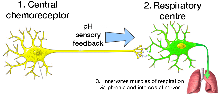 | |
Overview of neuronal pathway from central chemoreceptors to respiratory centre to respiratory muscles
Peripheral chemoreceptors
Peripheral receptors are located in the carotid and aortic bodies and are minor determinants of response to pCO2 accounting for approx 20% of response. Peripheral receptors respond to changes in pO2, pCO2 and H+
The carotid body nerve cells feedback via the glossopharyngeal nerve to the medulla of the brainstem. The aortic body, by contrast, is connected to the medulla via the vagus nerve.
Synergistic relationship of pO2 and pCO2 on ventilation
The major determinant of respiratory drive is an increase in pCO2 rather than a drop in pO2. Hypoxic drive usually only begins when pO2 falls below 8kPa. If a drop in pO2 is accompanied by a rise in pCO2 the effect on increasing ventilation is greater than the effects of either parameters alone. This is said to be the synergistic effect of pO2 and pCO2 on ventilation
The major determinant of respiratory drive is an increase in pCO2 rather than a drop in pO2. Hypoxic drive usually only begins when pO2 falls below 8kPa. If a drop in pO2 is accompanied by a rise in pCO2 the effect on increasing ventilation is greater than the effects of either parameters alone. This is said to be the synergistic effect of pO2 and pCO2 on ventilation
Respiratory centres & involuntary respiration
In addition to input from chemoreceptors the respiratory centres also receive sensory inputs form other parts of the brain and mechanoreceptors in other body tissues such as the lungs and GI tract.
 | |
There are 4 centres that effect ventilation change:
- Ventral respiratory group (VRG)
- Dorsal respiratory group (DRG)
- Apneustic centre
- Pneumotaxic centre
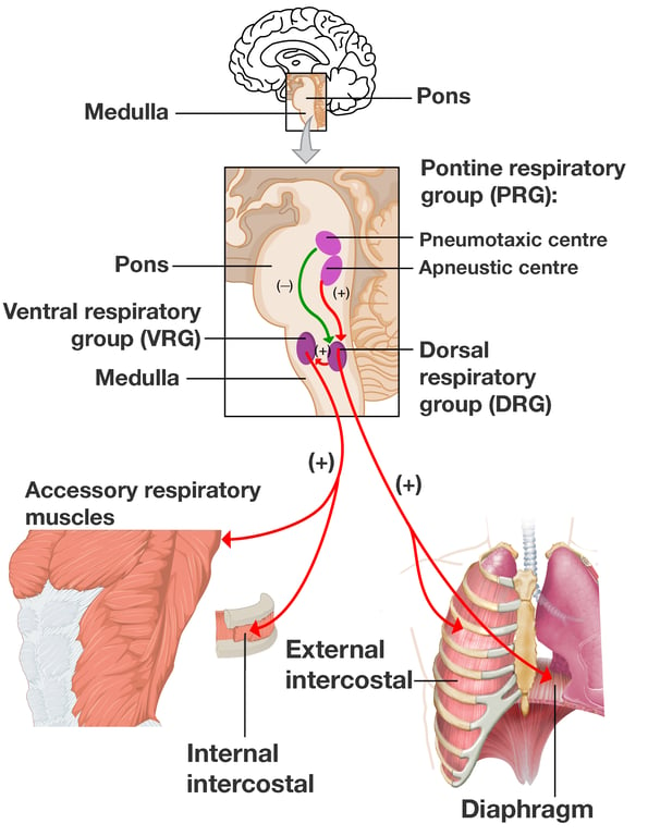 | |
Overview of respiratory control centres
The medulla oblongata is the primary respiratory control centre and is divided into dorsal and ventral neurone groups which have the following functions:
- Ventral respiratory group (VRG) stimulates expiratory movements.
- Dorsal respiratory group (DRG) stimulates inspiratory movements.
The pons contains the apneustic and pnuemotaxic centres which have the following functions:
- Apneustic centre stimulates inspiration for long and deep breaths, controls the intensity of breathing. It is inhibited by the stretch receptors of the pulmonary muscles at maximum depth of inspiration or by signals from the pnuemotaxic centre. It increases tidal volume.
- Pnuemotaxic centre sends signals to inhibit inspiration. Allows fine control of respiratory rate. Its signals limit the activity of the phrenic nerve and inhibits the signals of the apneustic centre. The pneumotaxic centre decreases depth and duration of inspiration resulting in decreased tidal volume and increased respiratory rate.
The apneustic and pnuemotaxic centers are antagonistic towards one another. Together these centres are sometimes collectively referred to as the pontine respiratory group (PRG)
Voluntary control of respiration
Voluntary respiration is any type of respiration that is under conscious control. Voluntary respiration is important for the higher functions that involve modulating air supply e.g. directed blowing (e.g. blowing out candles or performing valsalva), exaggerated inhalation or sniffing (e.g. when using inhaler device) or speaking and voice control.Voluntary respiration is initiated by the motor cortex within the cerebral cortex. Nerve impulses are sent to the diaphragm and intercostal muscles by the phrenic nerves, vagus nerves, and posterior thoracic nerves via the pyramidial tracts.
Voluntary control may be overridden by the autonomic system.
Note: The term pyramidal tracts refers to upper motor neurones that originate in the cerebral cortex and terminate in the spinal cord (corticospinal) or brainstem (corticobulbar)
Voluntary control of respiration is via cortical motor neurones in the pyramidial tracts