Respiratory physiology: Right to left shunts
Right to left shunts
A shunt is a passageway that allows movement of fluid from one part of the body to another.In the pulmonary circulation blood is typically moved from the right side of the heart to the left side of the heart via the lungs. This route allows the blood to become oxygenated.
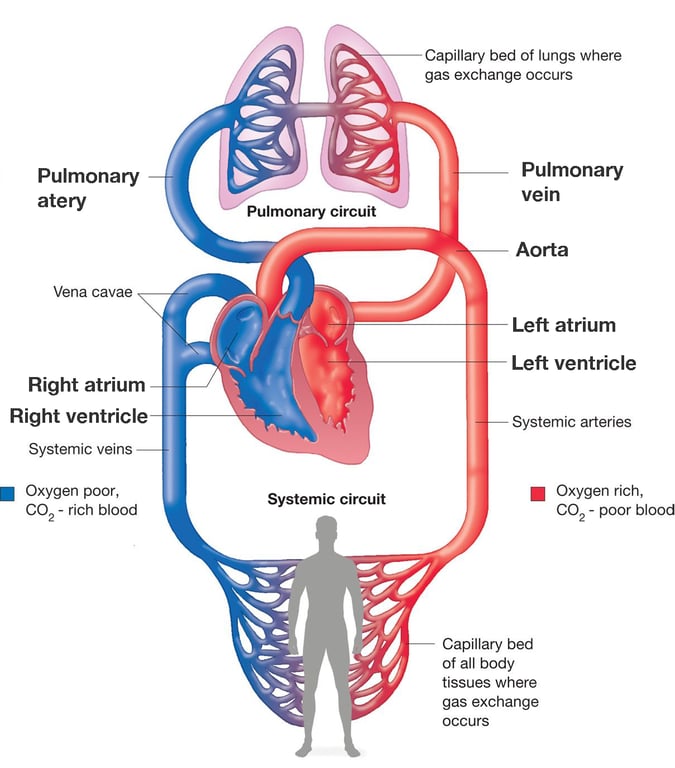 | |
Illustration of normal pulmonary circulation. In normal pulmonary circulation blood follows the following path: right atrium, right ventricle, pulmonary artery, capillary bed of lungs, pulmonary vein, left atrium, left ventricle then back to the systemic circulation via the aorta
Right to left shunts allow blood to bypass the pulmonary vasculature of the lungs and avoid oxygenation.
Physiological shunting
Some physiological shunting occurs in healthy individuals due to the following:Bronchial artery shunting: The bronchial arteries are branches of the thoracic aorta that anastomose with branches of the pulmonary arteries to supply the visceral pleura. The majority of this blood is returned via the pulmonary veins to the left side of the heart rather than the bronchial veins which drain to the azygos vein and subsequently the vena cava. As a result deoxygenated blood is returned to the left side of the heart.
Coronary artery shunting: Some of the blood supplied to the myocardium via the coronary arteries will drain via small cardiac veins (called Thebesian veins). Thebesian veins drain into both the right and left side of the heart. The blood draining into the left side is responsible for some physiological shunting
These physiological shunts do not usually have a deleterious effect as the deoxygenated shunted blood only accounts for around 1% of cardiac output.
Pathological shunting
Pathological right to left shunting may occur due to a number of conditions:- Persistent truncus arteriosus (minimal cyanosis)
- Transposition of great vessels
- Tricuspid atresia
- Tetralogy of Fallot
- Total anomalous pulmonary venous return
- Eisenmenger syndrome
- Re-opening of previously closed septal defects due to right heart hypertension
In pathological shunting a significant proportion of the blood arriving in the left ventricle for distribution to the systemic circulation may have bypassed the pulmonary vasculature. Shunted blood will have the following characteristics compared to non-shunted blood as a result of bypassing its opportunity for gas exchange to occur:
- Low pO2
- High pCO2
Detection of increased pCO2 and a subsequent compensatory increase in minute ventilation will typically prevent systemic hypercapnia developing until the shunt is greater than 50%. The hypoxemia owing to shunting is not responsive to increases in FiO2 because there is zero ventilation of the shunted blood (i.e. shunting could be considered physiologically comparable to a complete V/Q mismatch)