Bladder
On this page
Basic anatomy
The urinary bladder is a hollow muscular sac whose shape varies with filling. It can be thought of as pyramidal in shape. As can be seen from the diagrams below there are a number of key anatomical features.The bladder has an apex located superiorly and anteriorly. The apex points forward toward the upper part of the pubic symphysis.
The base is the posteroinferior surface of the bladder. It may also be referred to as the fundus. It contains the trigone.
The inferolateral surfaces extend inferiorly from the lateral borders and are limited posteriorly by the base whilst inferiorly they form the bladder neck. These surfaces are not covered by peritoneum.
The bladder neck is a constricted part of the urinary bladder where the inferolateral surfaces meet at the opening of the urethra. It is the most inferior part of the bladder. It is supported by puboprostatic ligaments (in males) or pubovesical ligaments (in females) and remains static when the bladder fills.
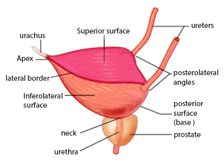 | |
Lateral view of the bladder. Superior surface highlighted in pink. Note the apex is the most anterior point of the bladder and points towards the pubic symphysis. It is connected to the umbilicus by the median umbilical ligament (a remnant of the urachus)
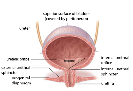 | |
Anterior view of the bladder. Note the peritoneum covers the superior surface and upper part of posterior surface only
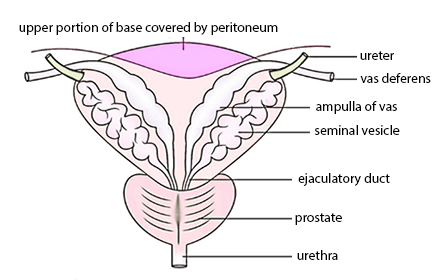 | |
Posterior view of the bladder base (fundus). Note the peritoneum covers the superior surface of the base
Trigone
The three orifices of the bladder are marked by the trigone. The two ureteric orifices and the internal urethral orifice can be thought of as the three points of the triangle and the area of the triangle is the trigone. The mucous membrane of trigone is always devoid of rugae.
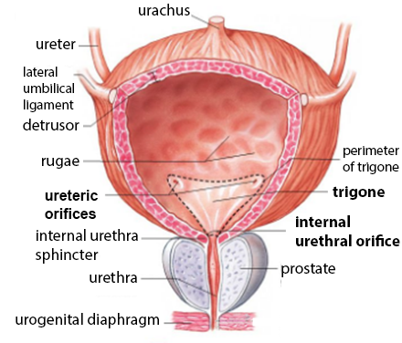 | |
Close up view highlighting the trigone
Innervation of the bladder
- Sympathetic supply via the hypogastric nerve (T11 - L2). It causes relaxation of the detrusor muscle and contraction of the sphincters.
- Parasympathetic supply via the pelvic nerve (S2-S4). It causes contraction of the detrusor muscle and inhibits urethral sphincter contraction thus stimulating micturition.
- The somatic nervous system allows for voluntary control over micturition via innervation of the external urethral sphincter. This is achieved via the pudendal nerve (S2-S4). It can cause it to constrict (storage) or relax (micturition).
- Sensory (afferent) nerves transmitting sensations of bladder fullness are conveyed to the spinal cord by the pelvic and hypogastric nerves whereas sensory input from the bladder neck and the urethra is carried in the pudendal and hypogastric nerves. The cell bodies of these neurons are at spinal levels of S2-S4 and T11-L2 spinal segments. Bladder pain is transmitted via the lateral spinothalamic tracts of the CNS.
Micturition
Micturition is a reflex action with can be modified by higher conscious control. The steps of the reflex are as follows:- Stretch receptors stimulated when bladder volume increases (typically >300ml)
- Afferent impulses fired which travel via S2-S4 and T11-L2 to the CNS
- The unconscious reflex part is processed by the lower micturition centre at S2-S4 level. The conscious control relies on impulses travelling up the spinal cord to the brainstem and back down again.
- The CNS spinal reflex triggers parasympathetic stimulation. Impulses travel in efferent fibres via S2-S4
- Detrusor contracts and urethral sphincters relax
Clinical significance
- Spinal micturition reflex reliant on S2-S4 being in tact.
- Cord lesions above S2: functioning bladder reflex but loss of conscious control and awareness of bladder filling. Bladder fills and empties every 1-4 hours
- Cord lesions below S2 (damage to micturition centre in S2-S4 spinal segments): Loss of parasympathetic outflow so detrusor doesn't contract. Bladder fills to capacity and overflows.