Root of the neck: subclavian artery and vein
Subclavian vein
Knowledge of the anatomy of the subclavian vein is required by the college and useful clinically given its use for central venous access.
Origin:
- axillary vein
- becomes subclavian vein as it passes behind junction of 1st rib and clavicle
- brachiocephalic vein
- forms brachiocephalic vein by merging with internal jugular vein behind sternoclavicular joint
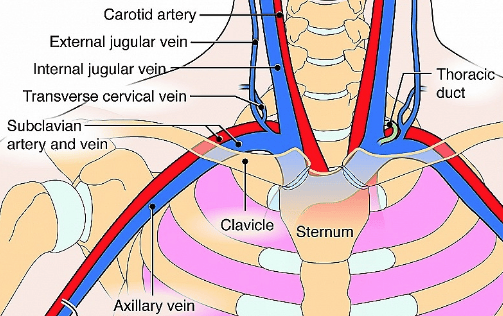 | |
Illustration showing arrangement of subclavian vessels
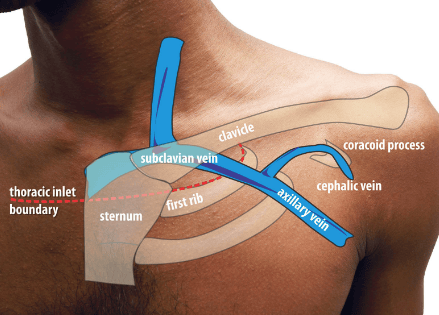 | |
surface markings of subclavian vein
Subclavian artery
Origin:
- Right: Brachiocephalic trunk behind sternoclavicular joint
- Left: Arch of aorta
Divided into 3 parts going from medial to lateral:
- 1st part: from the origin at the brachiocephalic trunk on the right and arch of aorta on the left to the medial border of scalenus anterior
- 2nd part: part posterior to scalenus anterior
- 3rd part: part lateral border of scalenus to outer border of first rib
- Terminates as axillary artery at outer border of 1st rib
Palpation
- The subclavian artery pulsation can be palpated 1 finger breadth above and parallel to the medial end of the clavicle.
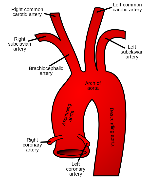 | |
Origins of the subclavian arteries
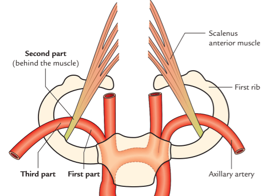 | |
Scalenus anterior dividing subclavian artery into 3 parts
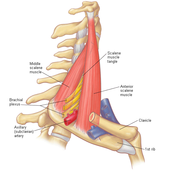 | |
Path of subclavian vessels in relation to scalenes