Pupillary light reflex
The pupillary light reflex involves testing both the direct and consensual pupil responses to light.
The direct reflex is seen when light is shone into the eye being observed and the consensual reflex is tested when light is shone into one eye but the contralateral eye is observed for pupil contraction.
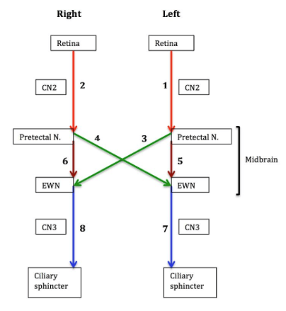
1. Light hits the retina and a nerve impulse is sent down the optic nerve to the midbrain.
2. Signal reaches the pretectal nucleus of the midbrain which responds by sending two efferent signals through crossed and uncrossed fibres (i.e. sends both ipsilateral and contralateral nerve impulses)
3. These signals travel via the Edinger-Westphal nuclei on each side of the midbrain to CN III on each side and finally onto the ciliary muscles of the pupil where the pupils constrict.
Diagrams illustrating schematic representations of pupil constriction
CN II Lesions
CN III Lesions
The direct reflex is seen when light is shone into the eye being observed and the consensual reflex is tested when light is shone into one eye but the contralateral eye is observed for pupil contraction.
Explanation
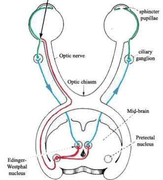
1. Light hits the retina and a nerve impulse is sent down the optic nerve to the midbrain.
2. Signal reaches the pretectal nucleus of the midbrain which responds by sending two efferent signals through crossed and uncrossed fibres (i.e. sends both ipsilateral and contralateral nerve impulses)
3. These signals travel via the Edinger-Westphal nuclei on each side of the midbrain to CN III on each side and finally onto the ciliary muscles of the pupil where the pupils constrict.
Diagrams illustrating schematic representations of pupil constriction
 | |
 | |
CN II Lesions
- When light is shone into side with CN II lesion produces an unresponsive pupillary light reflex on both sides
- With light is shone into the unaffected eye both pupils constrict.
CN III Lesions
- When light is shone into either eye there is lack of pupil constriction on the effected side (side with the lesion) and constriction on the normal side
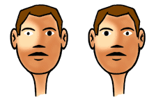 | |
The image on the left shows an example of light being shone into the normal side in a patient with a CN III lesion. Note appropriate direct constriction but the contralateral side remaining dilated indicating a CN III lesion on the consensual/contralateral side.
The image on the right shows an example of light being shone onto the side with the CN III lesion. Note the light stimulus must have successfully travelled down the right side via CN II to the midbrain. The midbrain has processed this signal but only the left pupil has received an efferent signal
The image on the right shows an example of light being shone onto the side with the CN III lesion. Note the light stimulus must have successfully travelled down the right side via CN II to the midbrain. The midbrain has processed this signal but only the left pupil has received an efferent signal