Head Injury
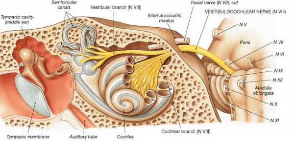
Diagram illustrating internal acoustic canal (meatus)
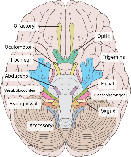
Image illustrating cranial nerve anatomy:
 | |
| Location (where cranial nerve exits skull) | Cranial nerve |
|---|---|
| cribriform plate | Olfactory nerve (I) |
| optic foramen | Optic nerve (II) |
| superior orbital fissure | Oculomotor (III) Trochlear (IV) Abducens (VI) Trigeminal V1 (ophthalmic) |
| Foramen rotundum | Trigeminal V2 (maxillary) |
| Foramen ovale | Trigeminal V3 (mandibular) |
| Stylomastoid foramen (via internal auditory canal) | Facial nerve (VII) |
| Internal auditory canal | Vestibulocochlear (VIII) |
| Jugular foramen | Glossopharyngeal (IX) Vagus (X) Accessory (XI) |
| Hypoglossal canal | Hypoglossal (XII) |
Image illustrating cranial nerve anatomy:
 | |
| Wikipedia | |