Platelets & haemostasis
Definition
Haemostasis is the mechanism that leads to cessation of bleeding from a blood vessel.Background
Haemostasis is a complex process that results in the formation of a blood clot and a fibrin mesh containing platelets and blood cells.Without effective haemostasis injury to the cardiovascular system would result in catastophic haemorrhage.
Platelets
Platelets (also known as thrombocytes) play a key role in homeostasis. They are formed in the bone marrow and released as fragments of megakaryocytes with each megakaryocyte producing 1,000-3,000 platelets during its lifetime.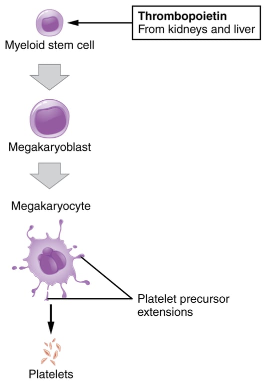 | |
Platelet production. Click on image to enlarge
The production of megakaryocytes and platelets is regulated by thrombopoietin, a glycoprotein hormone produced by the liver and kidney which stimulates the differentiation of stem cells into megakaryocytes.
A platelets lifespan is typically between 4 and 10 days. Platelets are primarily destroyed by phagocytosis by Kupfer cells. Approximately 2/3 of platelets circulate in the blood with the remaining 1/3 kept in reserve in the spleen. The RCEM defines a normal platelet count as 150 - 450 x 10*9/L
Key Points
- A platelets lifespan is typically between 4 and 10 days
- Formed in bone marrow via fragmentation of megakaryocytes
- Thrombopoietin regulates platelet production.
- Kupffer cells in the liver destroy old platelets, as well as the spleen.
Platelet structure
Platelets are small at 2-3 µm in maximum diameter. They have multiple surface receptors and contain granules. These granules contain mediators such as serotonin and ADP which are released via the platelets canalicular system to the exterior when they are activated.
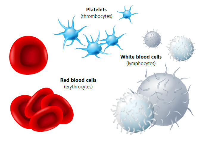 | |
Overview of platelet structure. Click on image to enlarge
Contents of platelet granules:
| Granule | Contents |
|---|---|
| Alpha granules |
|
| Delta or dense granules |
|
| Gamma granules (similar to lysosomes) |
|
| Lambda granules |
|
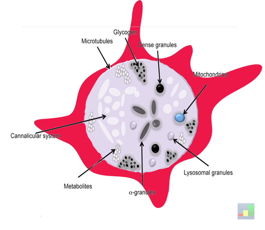 | |
Internal structure of platelet. Note that the external shape of the platelet changes from its inactive to activated state. Inactivated platelets resemble biconcave discs whereas an activated platelet demonstrates cell membrane projections covering their surface as shown above. Click on image to enlarge
Primary haemostasis
Primary haemostasis refers to the formation of the primary platelet plug. When a blood vessel is damaged the initial response is vasoconstriction to reduce haemorrhage followed by steps to seal the defect.The damaged vessel wall exposes collagen and tissue factor (TF). Exposed collagen binds to the plasma protein Von Willebrand factor (VWF).
Platelets have glycoprotein receptors that bind to VWF and integrins that bind to collagen. This results in platelets becoming tethered to the site of vessel wall damage. This step is referred to as
adhesion.
When platelets bind to these receptors they become
activated.
Platelet activation triggers a number of processes:- There is an increase in intracellular calcium
- Platelets change shape putting out pseudopodia
- Platelets produce thromboxane A2 (TXA2)
- TXA2 stimulates chemical release from platelet granules, namely 5-HT and ADP
- TXA2, 5HT and ADP cause vasoconstriction & other effects
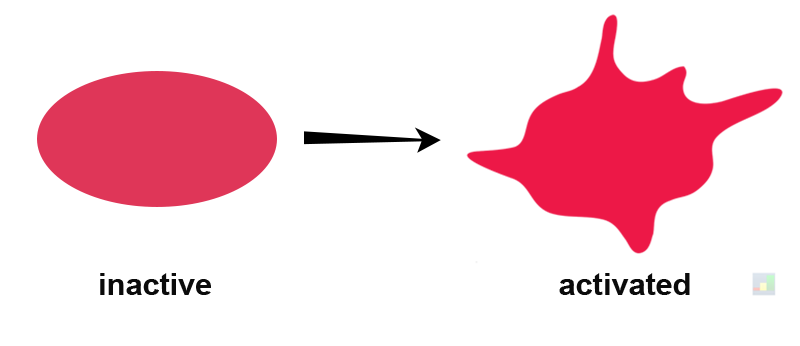 | |
Platelet activation triggers a change in platelet shape. Click on image to enlarge
Platelet degranulation results in the release of several chemical messengers including serotonin (5-HT) and ADP. The release of these chemical messengers causes a propagating effect as ADP activates more platelets by binding to purinergic receptors. ADP also activates fibrinogen receptors (GPIIb/IIIa) on the platelet surface causing platelets to bind to fibrinogen in plasma and cause them to become sticky and aggregate in a process known as platelet aggregation. Platelet aggregation results in the formation of the platelet plug.
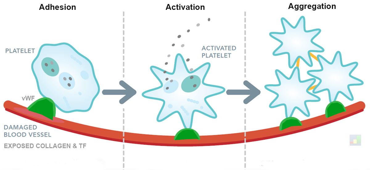 | |
Platelet adhesion, activation and aggregation simplified view. Click on image to enlarge
Clot formation
Following the formation of a platelet plug in primary haemostasis the next step is the formation of a fibrin mesh blood clot. This phase is often termed coagulation or secondary haemostasis.
Coagulation involves activation of several enzymes called clotting factors (a dozen or so plasma proteins that typically remain in an inactive state). The sequence of events has been referred to as the coagulation cascade in the past but the more current view is the cell based model of coagulation involving 4 overlapping steps of initiation, amplification, propagation and termination (see the video in the media links)
Both models ultimately lead to the formation of fibrin from inactive fibrinogen. These fibrin fibres form a mesh around the platelet plug to hold it in place.
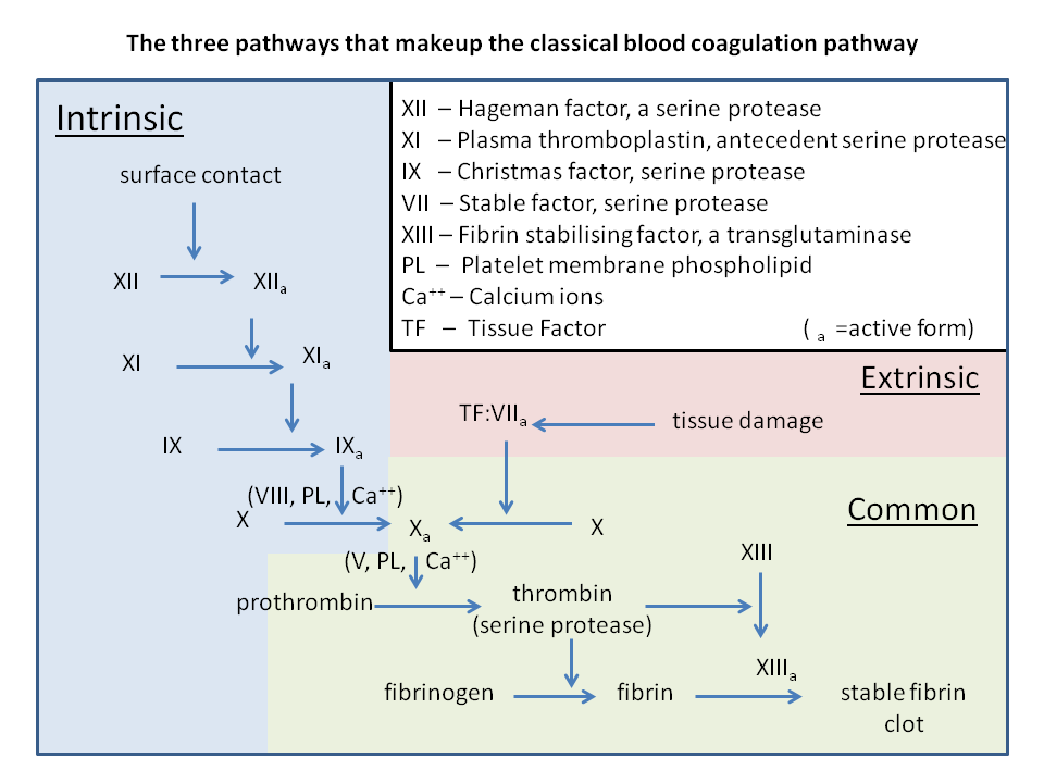 | |
An overview of the coagulation cascade. Click on image to enlarge
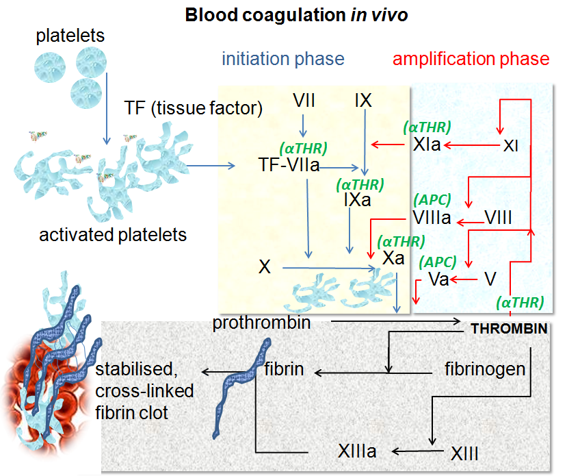 | |
An overview of the cell model of coagulation. Click on image to enlarge
The ultimate aim of coaglulation (secondary haemostasis) is to produce a large spike in thrombin levels that will cleave fibrinogen into fibrin and form a clot.
Inhibitors of haemostasis and fibrinolysis
As can be seen haemostasis involves a positive feedback loop. Positive feedback loops are unstable and require an inhibitor, in this case to prevent excessive coagulation.
These inhibitory mechanisms include:
- Undamaged endothelium produces prostacyclin and nitric oxide
- Plasma antithrombin inhibits thrombin, factor Xa and tenase (factor IXa:VIIIa complex)
- Thrombomodulin on undamaged endothelium binds thrombin and activates protein C. Protein C inactivates factors Va and VIIIa and tenase.
- Plasmin, produced by the activation of plasminogen by tissue plasminogen activator, breaks down fibrin clot in a process called fibrinolysis