Cardiac tamponade & pericardiocentesis
Candidates should note familiarity with the steps of this procedure are expected by the college and is highlighted in section 3 of the syllabus.
Complications of pericardiocentesis
It should be noted that pericardiocentesis is only used as a temporising measure pending thoracotomy. Recent articles published on the RCEM learning platform highlight the low success rate of pericardiocentesis and advise the procedure may also delay thoracotomy advocating against pericardiocentesis unless there is really no one capable of opening the chest.
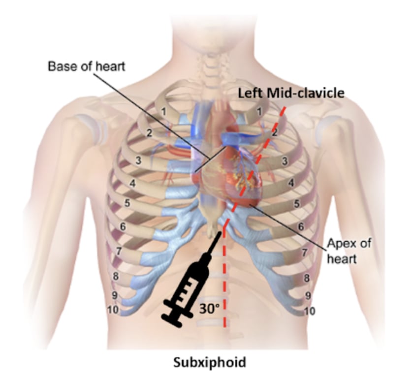 | |
The sub-xiphoid approach to pericardiocentesis. Click on image to enlarge
- Position patient in semi-reclined position with head of bed tilted at angle of 30-45 degrees
- Ensure ECG monitoring in place and skin cleansed with appropriate agent.
- Using long 18-22 G needle attached to syringe puncture the skin between xiphisternum and left costal margin (as shown in the diagram) at a 30º angle to the skin
- Needle is carefully advanced towards the tip of the left scapula whilst monitoring aspirant and ECG
- Once the right ventricle is reached and pericardial fluid is aspirated insert cannula and attach a 3 way tap. Further fluid may need to be removed to improve haemodynamics.
- Patients should have ECG monitoring
- Chest XRAY
- Ideally an echocardiogram
Complications of pericardiocentesis
- ST elevation or ventricular ectopics signal contact with ventricle
- Myocardial perforation
- Bleeding
- Pneumothorax
- Arrhythmia
- Acute pulmonary oedema (due to rapid drainage of pericardial fluid leading to excessive LV preload)
- Acute ventricular dilatation
It should be noted that pericardiocentesis is only used as a temporising measure pending thoracotomy. Recent articles published on the RCEM learning platform highlight the low success rate of pericardiocentesis and advise the procedure may also delay thoracotomy advocating against pericardiocentesis unless there is really no one capable of opening the chest.