Gastrointestinal physiology: Gallbladder
The gallbladder is a fibromuscular sac that lies on the posteroinferior surface of the right lobe of the liver (see diagram below)
Bile flows in and out of the gallbladder via the cystic duct. When the sphincter of Oddi closes in the interdigestive period pressure forces bile into the gallbladder where water is reabsorbed concentrating the bile.
When the sphincter of Oddi opens bile flows out of the cystic duct and into the small intestine via the common bile duct.
Following ingestion of food the gallbladder is stimulated to contract and empty by both neural and hormonal stimuli. The vagus nerve is the primary neural stimuli to contraction whilst CCK is the primary hormonal stimulant for the gallbladder to start emptying.
The release of CCK also promotes gallbladder contraction and emptying.
Hartmann's pouch is the outpouching of the gallbladder at the junction of neck and cystic duct. If gallstones impact here they may form Mucoceles.
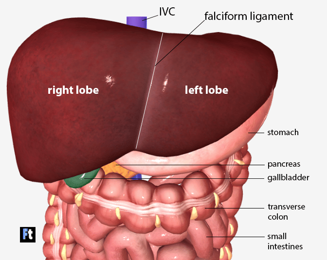 | |
Anterior view of the liver with gallbladder visible at inferior edge. Note its position anterior to the transverse colon
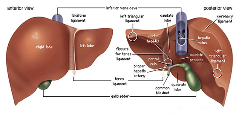 | |
Diagram illustrating position of gallbladder on posterior of liver
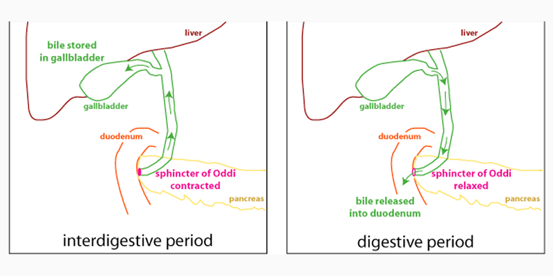 | |
Diagram illustrating gallbladder and bile flow
Bile flows in and out of the gallbladder via the cystic duct. When the sphincter of Oddi closes in the interdigestive period pressure forces bile into the gallbladder where water is reabsorbed concentrating the bile.
When the sphincter of Oddi opens bile flows out of the cystic duct and into the small intestine via the common bile duct.
Following ingestion of food the gallbladder is stimulated to contract and empty by both neural and hormonal stimuli. The vagus nerve is the primary neural stimuli to contraction whilst CCK is the primary hormonal stimulant for the gallbladder to start emptying.
The release of CCK also promotes gallbladder contraction and emptying.
Hartmann's pouch is the outpouching of the gallbladder at the junction of neck and cystic duct. If gallstones impact here they may form Mucoceles.
The gallbladder stores and concentrates bile. Concentration is via removal of non-essential solutes and water which leaves pigments and bile acids.
The process of concentration is driven by active transport of Na+ ions from gallbladder lumen into gallbladder epithelial cells then into the extracellular space. The sodium gradient drives the movement of water, bicarbonate and chloride ions intio the extracellular space.
The process of concentration is driven by active transport of Na+ ions from gallbladder lumen into gallbladder epithelial cells then into the extracellular space. The sodium gradient drives the movement of water, bicarbonate and chloride ions intio the extracellular space.