Ankylosing spondylitis: investigation and management
On this page
Ankylosing spondylitis is a HLA-B27 associated spondyloarthropathy. It typically presents in males (sex ratio 3:1) aged 20-30 years old.
Inflammatory markers (ESR, CRP) are typically raised although normal levels do not exclude ankylosing spondylitis.
HLA-B27 is of little use in making the diagnosis as it is positive in:
Plain x-ray of the sacroiliac joints is the most useful investigation in establishing the diagnosis. Radiographs may be normal early in disease, later changes include:
Spirometry may show a restrictive defect due to a combination of pulmonary fibrosis, kyphosis and ankylosis of the costovertebral joints.
The following is partly based on the 2010 EULAR guidelines (please see the link for more details):
Investigation
Inflammatory markers (ESR, CRP) are typically raised although normal levels do not exclude ankylosing spondylitis.
HLA-B27 is of little use in making the diagnosis as it is positive in:
- 90% of patients with ankylosing spondylitis
- 10% of normal patients
Plain x-ray of the sacroiliac joints is the most useful investigation in establishing the diagnosis. Radiographs may be normal early in disease, later changes include:
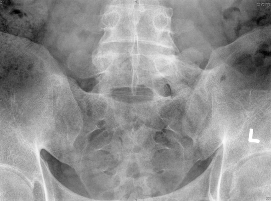
- sacroilitis: subchondral erosions, sclerosis
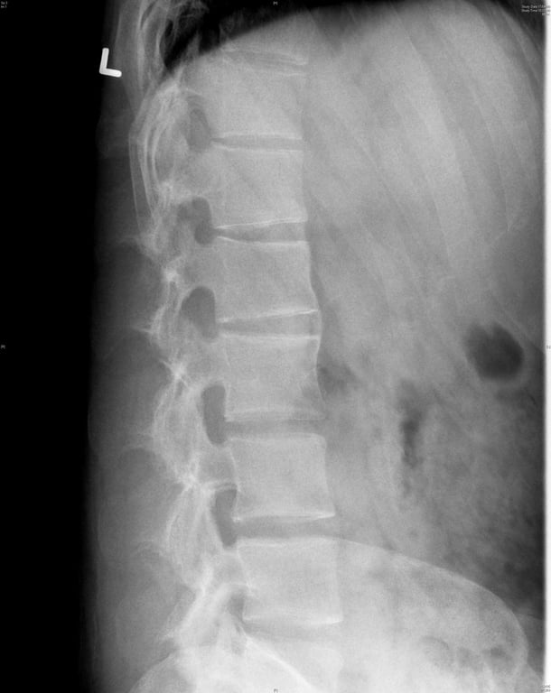
- squaring of lumbar vertebrae
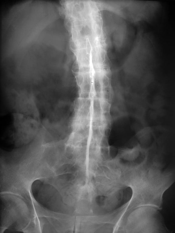
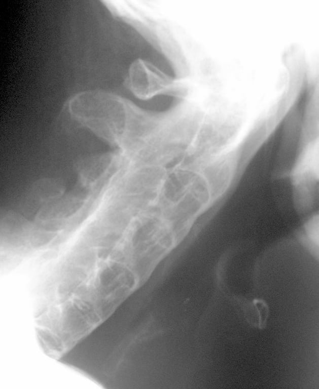
- 'bamboo spine' (late & uncommon)
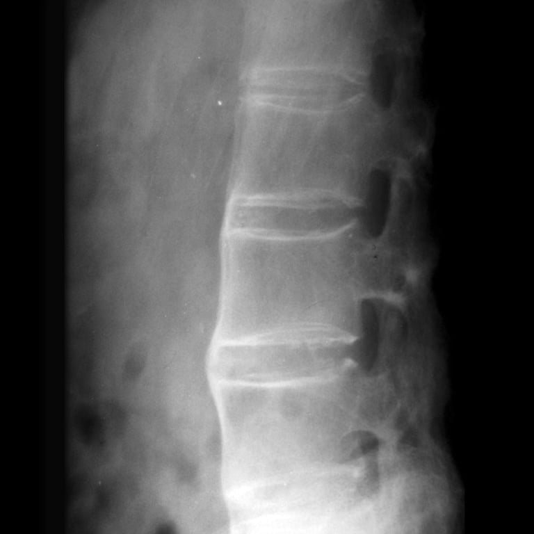
- syndesmophytes: due to ossification of outer fibers of annulus fibrosus
- chest x-ray: apical fibrosis
 | |
| © Image used on license from Radiopaedia | |
40-year-old male. There is typical appearance of bamboo spine with a single central radiodense line related to ossification of supraspinous and interspinous ligaments which is called dagger sign. Ankylosing is detectable in both sacroiliac joints
 | |
| © Image used on license from Radiopaedia | |
Ankylosing spondylitis with well formed syndesmophytes
 | |
| © Image used on license from Radiopaedia | |
Lateral cervical spine. Complete fusion of anterior and posterior elements in ankylosing spondylitis, so called bamboo spine
 | |
| © Image used on license from Radiopaedia | |
Fusion of bilateral sacroiliac joints. Sacroiliitis may present as sclerosis of joint margins which can be asymmetrical at early stage of disease, but is bilateral and symmetrical in late disease
 | |
| © Image used on license from Radiopaedia | |
Syndesmophytes and squaring of vertebral bodies. Squaring of anterior vertebral margins is due to osteitis of anterior corners. Syndesmophytes are due to ossification of outer fibers of annulus fibrosus
Spirometry may show a restrictive defect due to a combination of pulmonary fibrosis, kyphosis and ankylosis of the costovertebral joints.
Management
The following is partly based on the 2010 EULAR guidelines (please see the link for more details):
- encourage regular exercise such as swimming
- physiotherapy
- NSAIDs are the first-line treatment
- the disease-modifying drugs which are used to treat rheumatoid arthritis (such as sulphasalazine) are only really useful if there is peripheral joint involvement
- the 2010 EULAR guidelines suggest: 'Anti-TNF therapy should be given to patients with persistently high disease activity despite conventional treatments'
- research is ongoing to see whether anti-TNF therapies such as etanercept and adalimumab should be used earlier in the course of the disease