Gout: features
Gout is a form of inflammatory arthritis. Patients typically have episodes lasting several days when their gout flares and are often symptom free between episodes. The acute episodes typically develop maximal intensity with 12 hours/ The main features it presents with are:
Around 50% of first presentations affect the 1st metatarsophalangeal (MTP) joint. Attacks of gout affecting this area where historically called podogra. Other commonly affected joints include:
If untreated repeated acute episodes of gout can damage the joints resulting in a more chronic joint problem.
Radiological features of gout include:
Joint Aspirate:
- pain: this is often very significant
- swelling
- erythema
Around 50% of first presentations affect the 1st metatarsophalangeal (MTP) joint. Attacks of gout affecting this area where historically called podogra. Other commonly affected joints include:
- ankle
- wrist
- knee
If untreated repeated acute episodes of gout can damage the joints resulting in a more chronic joint problem.
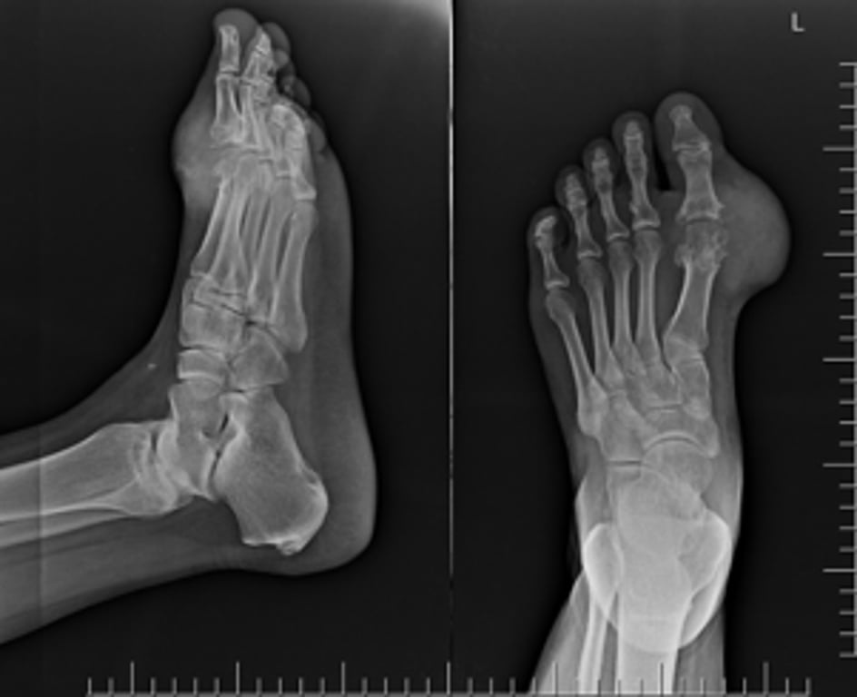
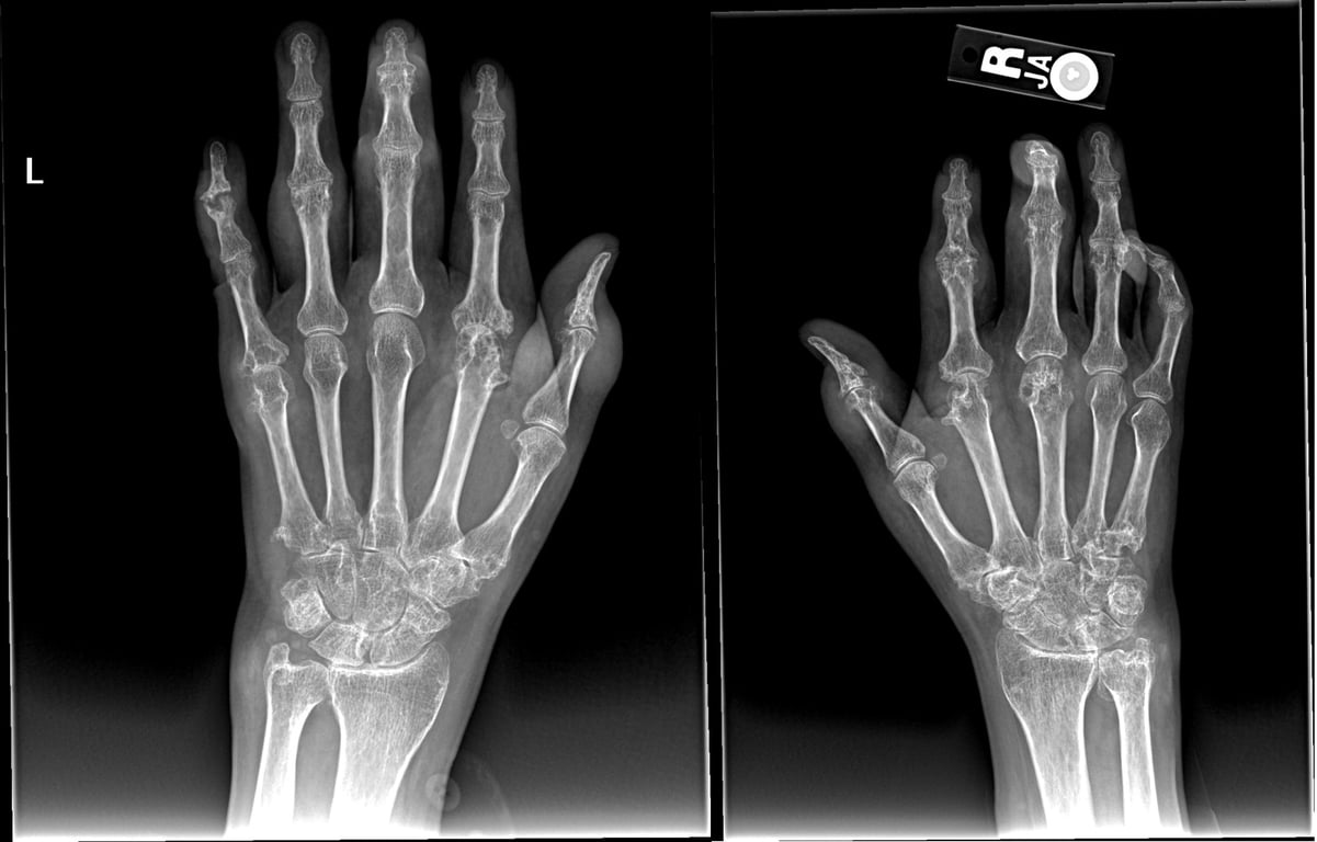
Radiological features of gout include:
- joint effusion is an early sign
- well-defined 'punched-out' erosions with sclerotic margins ina juxta-articular distribution, often with overhanging edges
- relative preservation of joint space until late disease
- eccentric erosions
- no periarticular osteopaenia (in contrast to rheumatoid arthritis)
- soft tissue tophi may be seen
Joint Aspirate:
- Negative birefringence
- Needle shaped blue crystals
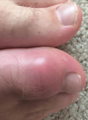 | |
Photograph of a patient with gout affecting the hallux.
 | |
| © Image used on license from Radiopaedia | |
X ray of a patient with gout affecting his feet. It demonstrates juxta-articular erosive changes around the 1st MTP joint with overhanging edges and associated with a moderate soft tissue swelling. The joint space is maintained.
 | |
| © Image used on license from Radiopaedia | |
X-ray of a patient with gout affecting his hands. There are multiple periarticular erosions bilaterally with adjacent large soft tissue masses and relatively preserved joint spaces. In the right hand, these findings are most prominent at the 1st interphalangeal, 2nd-4th proximal interphalangeal, 1st-3rd metacarpohalangeal and carpometacarpal joints. In the left hand, the findings are most prominent at the ulnar styloid, scapholunate joint, first and fifth carpometacarpal joints, second and fifth metacarpophalangeal joints and 1st interphalangeal joint.