Cardiovascular physiology: Heart sounds interpretation & the cardiac cycle
On this page
The first and second heart sounds are often referred to as S1 and S2 and should be audible in all individuals. Additional third (S3) and fourth (S4) heart sounds may be heard in some individuals and may be associated with some pathological conditions.
S1
S2
S3 (third heart sound)
S4 (fourth heart sound)
Heart sounds: sites of auscultation
Heart sounds: relation to cardiac cycle, ECG and CVP waveform
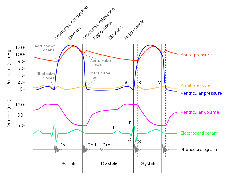
The diagrams below demonstrates the heart sounds in relation to the cardiac cycle, ECG and key pressures:
Diastole
The term diastole refers to the phase of the cardiac cycle where the chambers of the heart relax and undergo filling. When diastole is used on its own it is usually referring to ventricular diastole.
Diastole begins when the pulmonary and aortic valves (semilunar valves) close ending blood ejection from the ventricles. The ventricles can start to fill again when the atrioventricular valves (tricuspid and mitral) open.
Note the atrioventricular valves do not open immediately when the semilunar valves close, in fact there is a very short period of time after the semilunar valves close that both sets of valves are closed and this period is termed isovolumetric relaxation (the volume of blood within the ventricles cannot change due to the closed valves but the ventricles are starting to relax)
When the AV valves open the ventricles begin to fill. During the period that the ventricles are relaxing and filling the semilunar valves remain closed. Filling is initially a passive process with approximately 80-90% of filling occurring passively as the ventricles relax and expand. The second stage of filling occurs when the atria contract (atrial systole) and actively squeeze blood into the ventricles.
The amount of blood in the left ventricle at the end of filling is called the end-diastolic volume (EDV) and is typically around 120ml in healthy adults. When the ventricle contracts approximately 70ml is ejected (the stroke volume or SV). The ejection fraction can be calculated by SV/EDV
Heart sounds: summary
| Heart sound | Cardiac cycle phase | Cause |
|---|---|---|
| First (S1) | Start of ventricular systole | Closure of mitral and tricuspid (atrioventricular) valves |
| Second (S2) | Start of ventricular diastole | Closure of aortic and pulmonary (semilunar) valves |
| Third (S3) | During ventricular diastole | Blood striking left ventricle during passive ventricular filling |
| Fourth (S4) | Late ventricular diastole Atrial systole | Blood striking the left ventricle during active ventricular filling (atrial systole) |
S1
- closure of mitral and tricuspid valves
- soft if long PR or mitral regurgitation
- loud in mitral stenosis
S2
- closure of aortic and pulmonary valves
- soft in aortic stenosis
- splitting during inspiration is normal
S3 (third heart sound)
- caused by diastolic filling of the ventricle
- considered normal if < 30 years old (may persist in women up to 50 years old)
- heard in left ventricular failure (e.g. dilated cardiomyopathy), constrictive pericarditis (called a pericardial knock) and mitral regurgitation
S4 (fourth heart sound)
- may be heard in aortic stenosis, HOCM, hypertension
- caused by atrial contraction against a stiff ventricle
- in HOCM a double apical impulse may be felt as a result of a palpable S4
Heart sounds: sites of auscultation
| Valve | Site |
|---|---|
| Pulmonary valve | Left second intercostal space, at the upper sternal border |
| Aortic valve | Right second intercostal space, at the upper sternal border |
| Mitral valve | Left fifth intercostal space, just medial to mid clavicular line |
| Tricuspid valve | Left fourth intercostal space, at the lower left sternal border |
Heart sounds: relation to cardiac cycle, ECG and CVP waveform
The diagrams below demonstrates the heart sounds in relation to the cardiac cycle, ECG and key pressures:
 | |
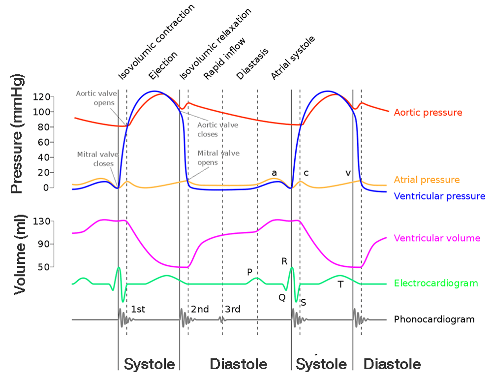 | |
Diagram illustrating the CVP waveform in relation to the cardiac cycle and ECG. Click on image to enlarge
Diastole
The term diastole refers to the phase of the cardiac cycle where the chambers of the heart relax and undergo filling. When diastole is used on its own it is usually referring to ventricular diastole.
Diastole begins when the pulmonary and aortic valves (semilunar valves) close ending blood ejection from the ventricles. The ventricles can start to fill again when the atrioventricular valves (tricuspid and mitral) open.
Note the atrioventricular valves do not open immediately when the semilunar valves close, in fact there is a very short period of time after the semilunar valves close that both sets of valves are closed and this period is termed isovolumetric relaxation (the volume of blood within the ventricles cannot change due to the closed valves but the ventricles are starting to relax)
When the AV valves open the ventricles begin to fill. During the period that the ventricles are relaxing and filling the semilunar valves remain closed. Filling is initially a passive process with approximately 80-90% of filling occurring passively as the ventricles relax and expand. The second stage of filling occurs when the atria contract (atrial systole) and actively squeeze blood into the ventricles.
The amount of blood in the left ventricle at the end of filling is called the end-diastolic volume (EDV) and is typically around 120ml in healthy adults. When the ventricle contracts approximately 70ml is ejected (the stroke volume or SV). The ejection fraction can be calculated by SV/EDV
Diastole Key Points:
- During diastole 80-90% of ventricular filling is passive. 10-20% of filling is secondary to atrial contraction (systole)
- In a healthy adult EDV is approximately 120ml whilst stroke volume (SV) is 70 ml
- End diastolic pressure is approximately 4-7mmHg
- AV valves open
- Semilunar valves remain closed
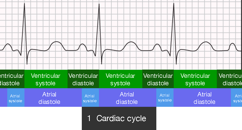 | |
A cardiac cycle begins with atrial systole (which corresponds to the P-wave on the ECG) and ends at the start of the next P-wave. Click on image to enlarge
Systole
During systole the ventricles contract. The rising pressure first closes the atrioventricular valves which prevents backflow of blood to the atria. For a short period of time both the AV and semilunar valves are closed - this is termed isovolumetric contraction as the blood volume within the ventricle cannot change but the pressure rises. The rising ventricular pressure eventually forces the semilunar valves to open and blood is ejected into the pulmonary artery and aorta.The cardiac cycle starts with atrial contraction (atrial systole) which corresponds to the P-wave on the ECG