Muscle contraction: excitation-contraction coupling
On this page
Excitation-contraction coupling
Skeletal muscle contraction may be broken down into excitation and contraction phases. Excitation-contraction coupling in the skeletal muscle refers to the sequence of events through which nerve fibres stimulate skeletal muscle fibres causing them to contract.Excitation sequence in skeletal muscle
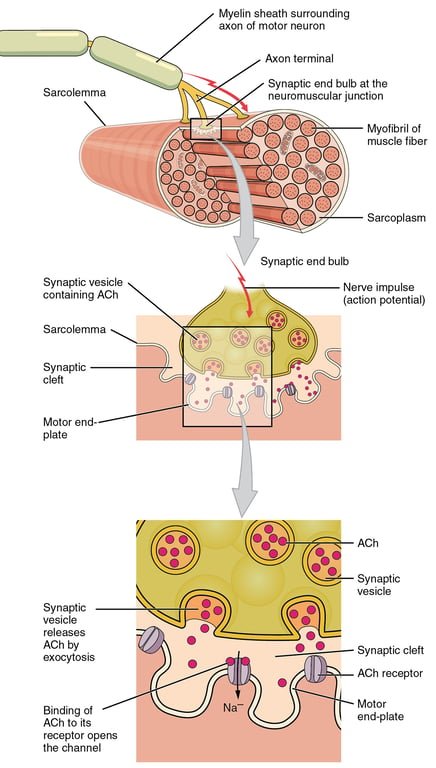
- Action potential reaches the end bulb of the nerve at the neuromuscular junction causing the polarity inside the end bulb to change (becomes positive). This opens calcium channels on the nerve end bulb resulting in calcium ion influx through voltage-gated calcium channels.
- Calcium influx causes the release of acetylcholine form vesicles into the synapatic space or cleft via exocytosis.
- Acetylcholine crosses the synapse and binds to nicotinic acetylcholine receptors of the muscle cell. This causes opening of ligand gated sodium channels on the muscle and an influx of sodium, depolarising the muscle membrane.
- Depolarisation spreads through the muscle primarily via T-tubules
- Depolarization activates L-type voltage-dependent calcium channels (dihydropyridine receptors) in the T-tubule membrane, which are close to calcium-release channels (ryanodine receptors) in the adjacent sarcoplasmic reticulum
- Sarcoplasmic reticulum releases calcium ions
 | |
click on image to enlarge
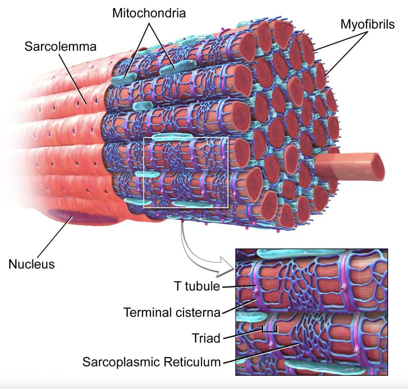 | |
Diagram illustrating skeletal muscle highlighting sarcolemma. click on image to enlarge
Contraction sequence in skeletal muscle
- The increased intracellular Ca2+ ion concentration within the muscle is the start of the contraction sequence.
- Ca2+ binds to troponin (found on actin-containing thin filaments) causing a conformational change, allowing tropomyosin to move, unblocking the binding sites
- Myosin head binds to the newly released binding site then pulls the actin filament by tilting its head (the power stroke), pulling the Z bands towards each other.
- The power stroke releases the bound ADP and allows ATP to bind to myosin which causes the myosin-actin crossbridge to dissociate.
Terminology notes
| Component | Notes |
|---|---|
| Sarcomere | Basic unit of muscles that gives skeletal and cardiac muscles their striated appearance. Segment between two adjacent Z-lines |
| I-band | Zone of thin filaments that is not superimposed by thick filaments |
| A-band | Contains the entire length of a single thick filament |
| H-zone | Zone of the thick filaments that is not superimposed by the thin filaments |
| M-line | Middle of the sarcomere, cross-linking myosin |
| Sarcoplasmic reticulum | Releases calcium ion in response to depolarization |
| Actin | Thin filaments that transmit the forces generated by myosin to the ends of the muscle |
| Myosin | Thick filaments Binds to thin filament |
| Titin | Connects the Z-line to the thick filament, altering the structure of tropomyosin |
| Tropomyosin | Covers the myosin-binding sites on actin |
| Troponin-C | Binds with calcium ions |
| T-tubule | Is an invagination of the sarcoplasmic reticulum Helps co-ordinate muscular contraction |
The table below shows the types of skeletal muscle fibres (type II fibres are simplified into one category):
| Type I | Type II | |
|---|---|---|
| Contraction time | Slow | Fast |
| Colour | Red (due to presence of myoglobin) | White (due to absence of myoglobin) |
| Main use | Sustained force | Sudden movement |
| Major fuel | Triglycerides | ATP |
| Mitochondrial density | High | Low |
Mnemonic for type I fibres - 'One Slow, Fat, Red Ox'