Kidneys: renal anatomy
On this page
Surface anatomy of the kidneys
- Posteriorly the kidneys occupy from T12 to L3 levels.
- The hilum is typically found at L1 level i.e. in the transpyloric plane
- The right kidney is lower than the left kidney
- The kidneys move downwards by 2.5 cm on inspiration
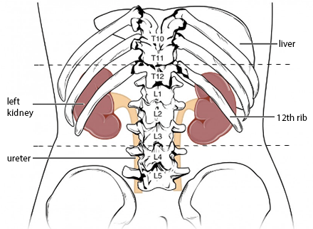 | |
illustration of the surface anatomy of the kidney. Note the right kidney sits lower than the left kidney as it is pushed down by the liver.
Layers covering the kidney
From deep -> superficilal:- Fibrous capsule
- Perirenal fat
- Renal fascia
- Pararenal fat
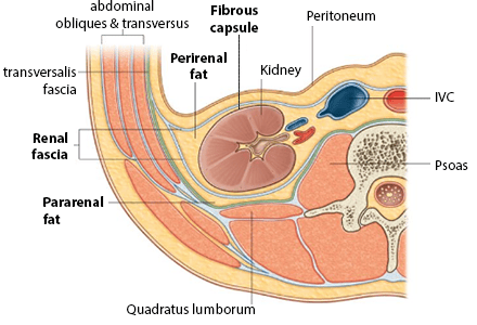 | |
illustration of the layers covering the kidney. The renal fascia is important as a ruptured kidney abscess will be contained within the fascia. Pus may point above the iliac crest posteriorly
Arterial supply to the kidneys
- The renal arteries arise directly from the abdominal aorta at L1-L2 level posterior to the pancreas.
- Renal arteries are end arteries. Obstruction leads to infarction.
- Renal arteries divide into anterior and posterior branches then segmental branches before the segmental arteries enter the hilum which typically lies at L1
- Renal arteries divide as below:
renal artery → segmental arteries→ lobar arteries → interlobar arteries → arcuate arteries
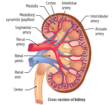 | |
illustration of blood supply to the kidney
Lymph drainage of the kidneys
- Para-aortic nodes
Innervation of the kidneys
- From renal plexus which is formed by the coeliac plexus, sympathetic trunk and spinal nerves
- Sensory afferents from the renal pelvis and ureter travel to T11-L2 hence the flank, loin and groin pain felt in renal colic. As the genitofemoral nerve originates from the upper L1-2 segments of the lumbar plexus pain may also be felt down the leg as pain is transmitted via the femoral branch of the genitofemoral nerve.
Anatomical relations of the kidneys
Right kidney
| Direct contact or Layer of peritoneum in-between | Anterior to kidney | Posterior to kidney |
|---|---|---|
In direct contact
|
Left kidney
| Direct contact or Layer of peritoneum in-between | Anterior to kidney | Posterior to kidney |
|---|---|---|
In direct contact
|
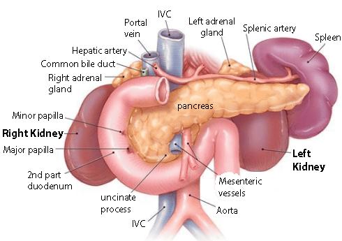 | |
illustration of the kidneys and relations to nearby organs